Browse by specialty
- Anesthesia and Perioperative
- Cardiology & Cardiovascular System
- Clinical Pharmacology
- Dermatology
- Diagnostic Imaging
- Emergency Medicine
- Endocrinology
- Ethical, Legal, and Organizational Medicine
- Family Medicine
- Gastroenterology
- General Surgery
- Geriatric Medicine
- Gynecology
- Haematology
- Infectious Disease
- Nephrology
- Neurology
- Neurosurgery
- Obstetrics
- Ophthalmology
- Orthopedics
- Otolaryngology
- Pediatrics
- Plastic Surgery
- Population and Community Health
- Psychiatry
- Respirology
- Rheumatology
- Urology
Ophthalmology
Full-Colour Atlas
Age Related
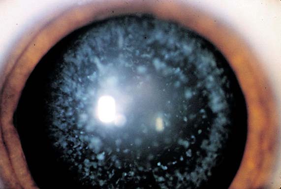
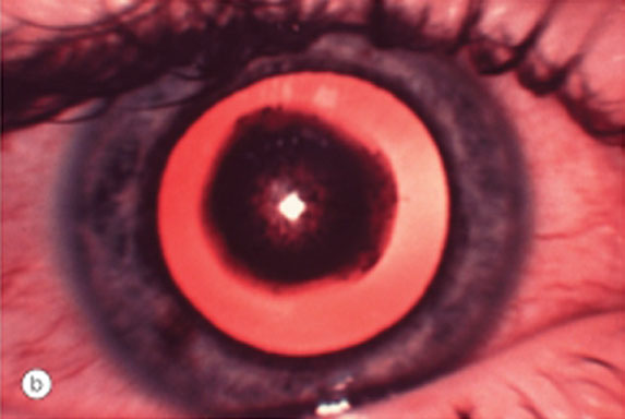
Cataract (Age related cataract)
Posterior subcapsular cataract type seen on retroillumination. (Courtesy of Elsevier)
Corneal Abrasion
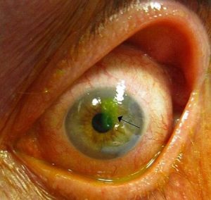
(Source: James Heilman, MD and licensed under the Creative Commons Attribution-Share Alike 3.0 Unported license)
Available from: https://commons.wikimedia.org/wiki/File:Human_cornea_with_abrasion_highlighted_by_fluorescein_staining.jpg
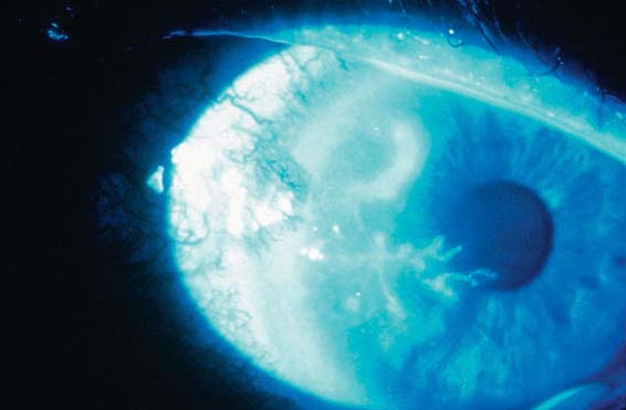
Herpes Simplex Keratitis
Irregular dendritic (branch-like) lesion of corneal epithelium stained with fluorescein.
Viral Conjunctivitis No. 1
Adenoviral conjunctivitis with conjunctival injection. (Courtesy of Department of Ophthalmology, University of Toronto)
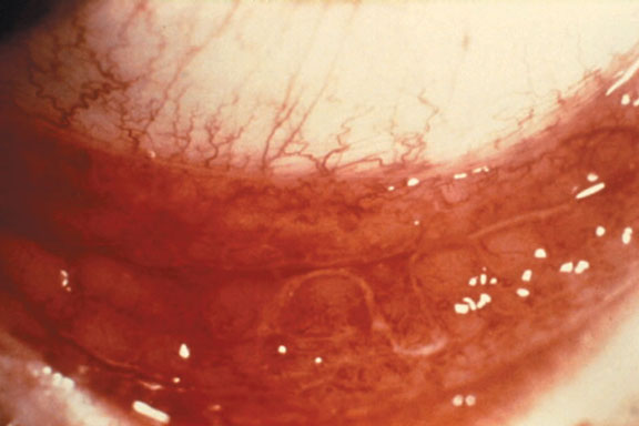
Viral Conjunctivitis No. 2
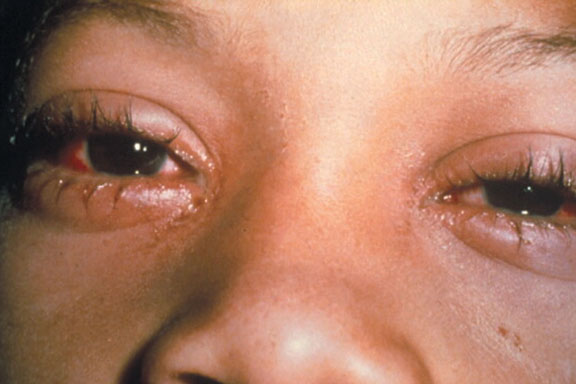
Adenoviral conjunctivitis with lid swelling, conjunctival injection and tearing. (Courtesy of Department of Ophthalmology, University of Toronto)
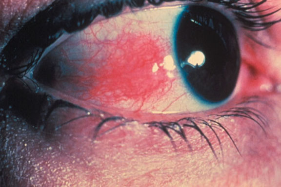
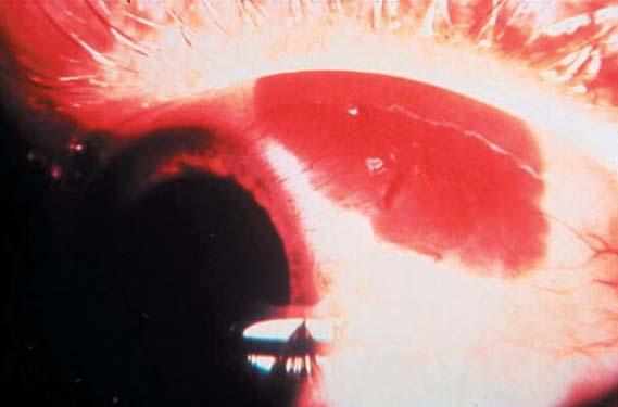
Subconjunctival Haemorrhage
Subconjunctival haemorrhage as evidenced by a bright red colour. (Courtesy of Department of Ophthalmology, University of Toronto)
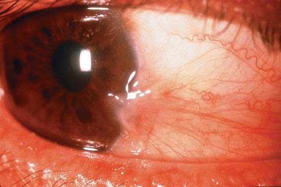
Pterygium
Pterygium extends onto the cornea (Courtesy of Department of Ophthalmology, University of Toronto)
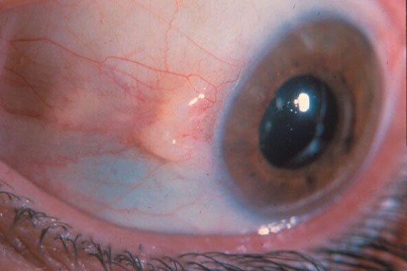
Pinguecula
Pingueculum is degenerative collagen within the interpalbebral fissure. (Courtesy of Department of Ophthalmology, University of Toronto)
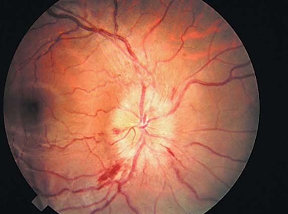
Papilledema
Elevated congested disc with indistinct margins, flameshaped hemorrhages, and dilated tortuous vessels.
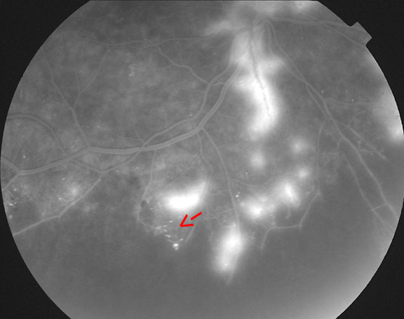
Neovascularization of the Disc
(NVD) Early fluorescein angiography image of the eye in image OP35 demonstrating areas of vascular leakage near the disc. 1. NVD; 2. Vascular leakage
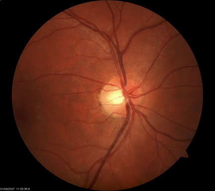
Neovascularization Elsewhere
(NVE) Fluorescein angiography image of the eye in image OP35 showing areas of vascular leakage.
Titled Disc No. 1
Titled discs are associated with high myopia or moderate oblique myopic astigmatism. Although titled discs have no systemic or neurologic association, the visual field may show bitemporal field defects usually confined to the superior quadrant. Patients with severe myopia should be evaluated by a retina specialist at least once yearly with scleral depression and indirect ophthalmoscopy due to risk of retinal tear and subsequent detachment.
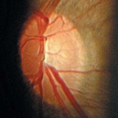
Tilted Disc No. 2
Titled discs are associated with high myopia or moderate oblique myopic astigmatism. Although titled discs have no systemic or neurologic association, the visual field may show bitemporal field defects usually confined to the superior quadrant. Patients with severe myopia should be evaluated by a retina specialist at least once yearly with scleral depression and indirect ophthalmoscopy due to risk of retinal tear and subsequent detachment.
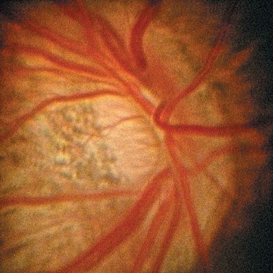
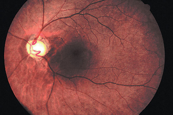
Optic Disc Enlargement No. 1
Optic Disc Enlargement, thinning (or notching) of neuroretinal rim usually beginning inferiorly and deepening of optic cup. Notching thinning of neuroretinal rim tends to occur, inferiorly, then superiorly, then temporally, and nasal is the last on to be affected. (Courtesy of 2003 Dr. Yan)
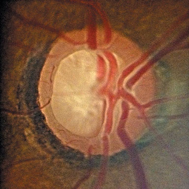
Optic Disc Enlargement No. 2
Slight thinning of neuroretinal rim inferiorly and temporally, deepening of cup, development of pallor (late finding of glaucoma).
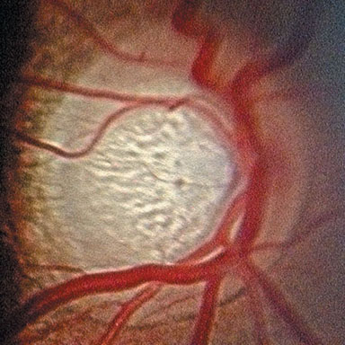
Optic Disc Enlargement No. 3
Optic Disc Enlargement, thinning (or notching) of neuroretinal rim usually beginning inferiorly and deepening of optic cup. Notching thinning of neuroretinal rim tends to occur, inferiorly, then superiorly, then temporally, and nasal is the last on to be affected. (Courtesy of 2003 Dr. Yan)

Chemical Burns

Leukocoria

Ophthalmia Neonatorum
Chemical Burns
Chemical Burns
Please refer to the following website for images of Chemical Burns (Figures A, C, and D) under Diagnosis: https://eyewiki.org/Chemical_(Alkali_and_Acid)_Injury_of_the_Conjunctiva_and_Cornea
Vitreous
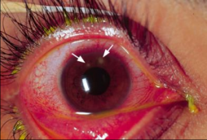
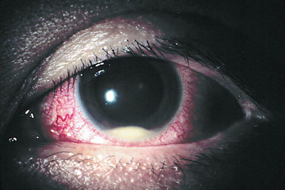
Endophthalmitis with Hypopyon
Prominent layer of purulent material in inferior aspect of anterior chamber. Note corneal edema and conjunctival injection.
Scleritis
Scleritis with diffuse involvement on the deep episcleral vessals. (Courtesy of Department of Ophthalmology, University of Toronto)
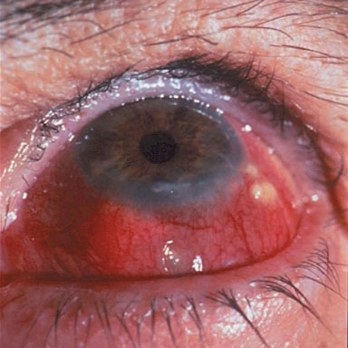
Episcleritis
Episcleritis with sectorial injection of the conjunctiva and episcleral tissue. (Courtesy of Department of Ophthalmology, University of Toronto)
