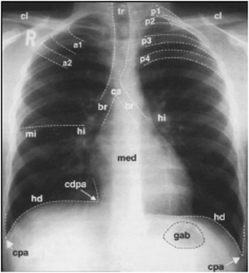
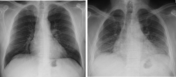
Normal

PA Film

Normal PA film of a male. Note the right and left clavicles (cl), posterior (p1-4) and anterior (a1-2) ribs, right and left costophrenic angles(cpa), right cardiophrenic angles (cdpa), right and left hemidiaphragms (hd), gastric air bubble (gab), trachea (tr), right and left mainstem bronchi (br), mediastinal shadow (med), carina (ca), and right and left hila (hi). The normal position of the minor fissure (mi) is also indicated.

PA Film

Normal – AP

Because the AP projection positions the heart and mediastinum further from the x-ray film than the PA projection, these structures appear magnified with the AP view. Other clues that you are looking at an AP projection include clavicles that project above the lung apices and scapulae that project over the lung fields.

Normal - AP

Nipple Shadows

Nipple shadows can often mimic pulmonary nodules. Clues to nipples being the source of the apparent nodules on the lateral radiograph, bilaterally and “lesions” whose inferior and lateral borders appear sharper than their superior and medial margins. Confirmation can be obtained by repeating the study with nipple markers or obtaining a different projection.

Nipple Shadows

Mediastinum

Components of the PA mediastianl shadow (A) include SVC (1), ascending aorta (2), RA (3), IVC (4), aortic arch (5), pulmonary trunk (6), LA appendage (7), and LV (8).
Mediastinal compartments on the lateral film (B) include: superior (S), anterior (A), middle (M) and posterior (P) compartments.

Mediastinum

Lateral Film

This is a normal lateral film of a female patient. Note the spine (sp), anterior costophrenic angle (acpa), gastric air bubble (gab), trachea (tr), left mainstem bronchus (lbr), right mainstem bronchus (rbr), aortic arch (ao), anterior/retrosternal (acs) and posterior/retrocardiac (pcs) clear spaces, and breast shadow (m)

Lateral Film

Inspiration and Expiration Films

On a film taken in full inspiration, the right hemidiaphragm should project over the 6th anterior interspace or 10th rib posteriorly. Films taken without a full inspiration are described as having a “poor inspiratory result”. This may result from a poor inspiratory effort or any other condition that prevents full inspiration.

This patient’s chest x-ray is normal in full inspiration. In relative expiration, the cardiac silhouette appears enlarged and the pulmonary vasculature appears crowded and indistinct. This appearance is easily mistaken for pulmonary edema

Inspiration and Expiration Films