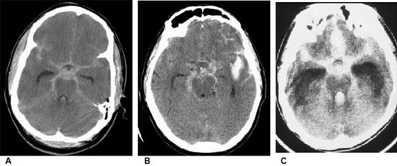
Subarachnoid Hematoma
Image A. Axial CT image with blood filling the suprasellar cistern, ambient cisterns, interhemispheric fissure, and sylvian fissures in the classic star pattern.
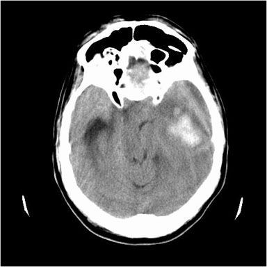
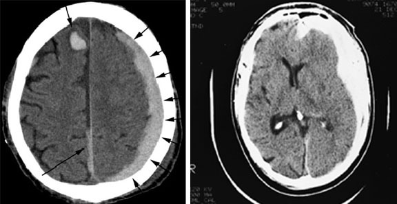
Image B. Subarachnoid hemorrhage and aneurysm. Axial non-contrast CT image showing a SAH and right MCA aneurysm with mild hydrocephalus seen by the prominent temporal horns of the lateral ventricles.
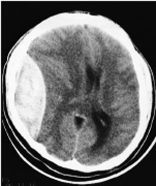
Image C. CT without contrast showing blood in basal and suprasellar cisterns, interhemispheric and Sylvian fissures
[Courtesy of Dr. J. Spears]