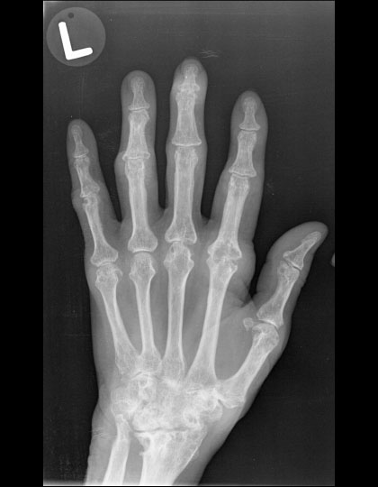
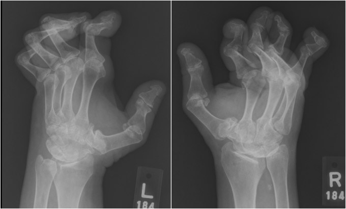
Figure – 14
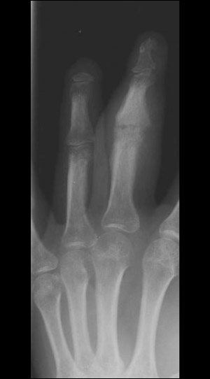
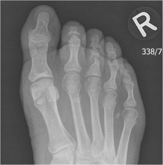
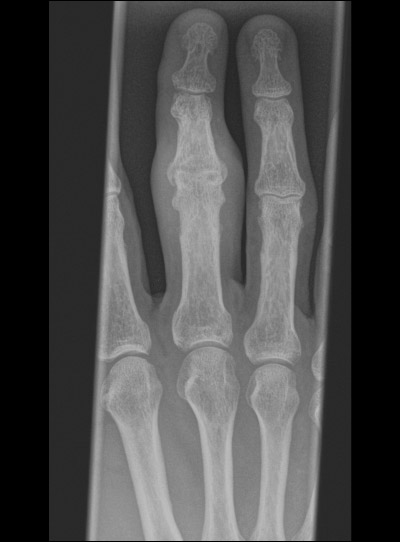
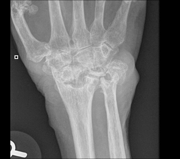
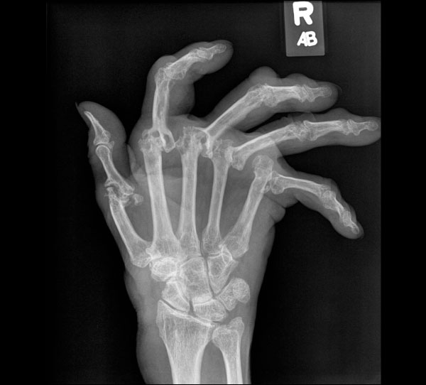
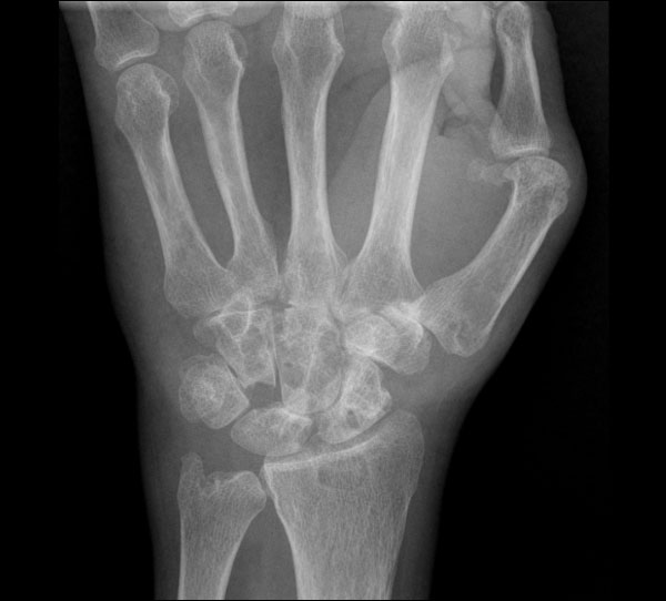
Left/right hand radiograph in a patient with osteoarthritis (OA), which shows joint space narrowing of the DIPs, osteophytes, and bone remodelling. These radiographic features are typical in mild to severe OA.
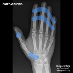
Case courtesy of Dr. Craig Hacking, Radiopaedia.org, rID: 69224