Browse by specialty
- Anesthesia and Perioperative
- Cardiology & Cardiovascular System
- Clinical Pharmacology
- Dermatology
- Diagnostic Imaging
- Emergency Medicine
- Endocrinology
- Ethical, Legal, and Organizational Medicine
- Family Medicine
- Gastroenterology
- General Surgery
- Geriatric Medicine
- Gynecology
- Haematology
- Infectious Disease
- Nephrology
- Neurology
- Neurosurgery
- Obstetrics
- Ophthalmology
- Orthopedics
- Otolaryngology
- Pediatrics
- Plastic Surgery
- Population and Community Health
- Psychiatry
- Respirology
- Rheumatology
- Urology
Gynecology
Full-Colour Atlas
Adenomyosis – MRI
MRI image of adenomyosis.
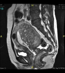
Adenomyosis MRI Image (Case courtesy of Dr. Varun Babu, Radiopaedia.org, rID: 43504)
“The most easily recognized feature is a thickening of the junctional zone ≥12 mm, either diffusely or focally (normal junctional zone thickness is up to ~5 mm)”. (1)
(1) Gaillard F, Thibodeau R, Liao A, et al. Adenomyosis. Reference article, Radiopaedia.org (Accessed on 17 May 2024). https://doi.org/10.53347/rID-10171.
Adenomyosis – Ultrasound
Ultrasound image of adenomyosis.
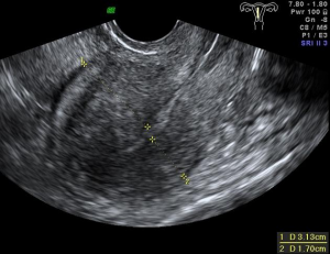
Adenomyosis Ultrasound Image (Case courtesy of Dr. Alexandra Stanislavsky, Radiopaedia.org, rID: 13726)
“MUSA features typical of a uterus with adenomyosis include an enlarged globular uterus, asymmetrical thickening of the myometrium, myometrial cysts, echogenic subendometrial lines and buds, hyperechogenic islands, fan-shaped shadowing, an irregular or interrupted junctional zone and translesional vascularity on colour Doppler ultrasound examination.”(1)
(1) Van den Bosch, T., de Bruijn, A.M., de Leeuw, R.A., Dueholm, M., Exacoustos, C., Valentin, L., Bourne, T., Timmerman, D. and Huirne, J.A.F. (2019), Sonographic classification and reporting system for diagnosing adenomyosis. Ultrasound Obstet Gynecol, 53: 576-582. https://doi.org/10.1002/uog.19096
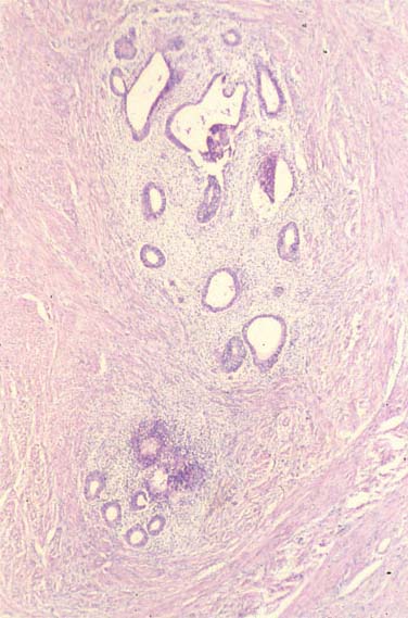
Adenomyosis
Microscopic endometrial stroma and glands present deep within the myometrium. (Courtesy of Dr. I. Zberiranowski)
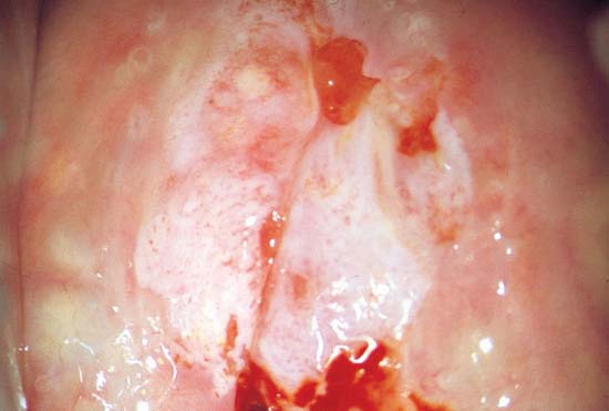
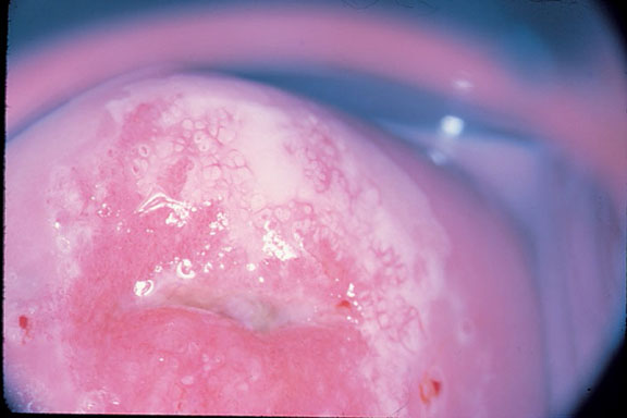
Squamous Intra-epithelial
Low-grade squamous intra-epithelial lesion stained with acetic acid. (Courtesy of Dr. G. Likrish)
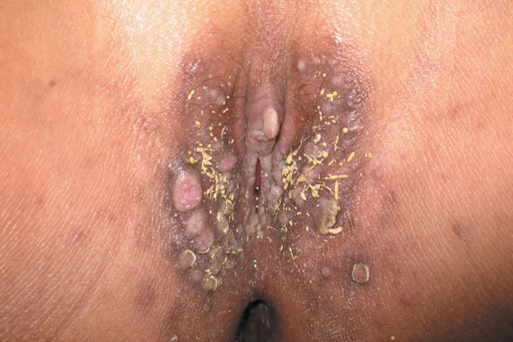
Condyloma Acuminate – External Genitalia
Condyloma Acuminate (“Genital Warts”)
Soft cauliflower-like, skin-coloured masses in clusters; associated with human papilloma virus (HPV). (Courtesy Dr. S. Kives)
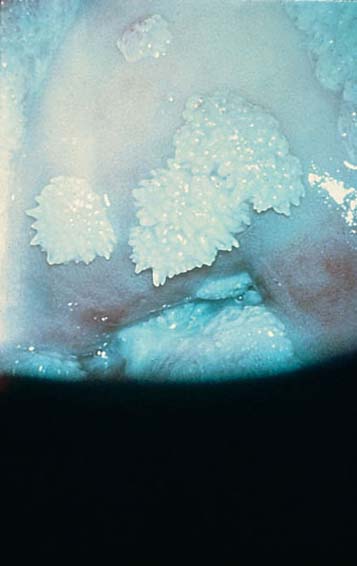
Condyloma Acuminate – Cervix
View of the cervix. Range from pinhead to papules. (Courtesy of Dr. W. Chapman)
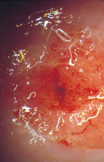
Ectropion
Eversion of cervical canal, with columnar epithelium farther outside the external os of the cervix. (Courtesy of Dr. G. Likrish)
Endometrioma
Cystic mass on ovary arising from ectopic endometrial tissue. Commonly referred to as a “chocolate cyst”.
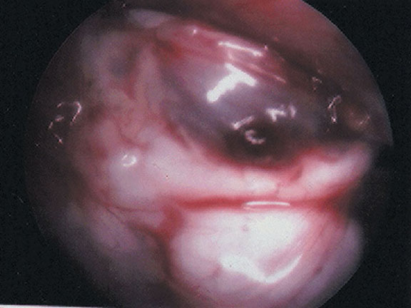
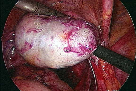
Female Anatomy – Normal
Laparoscopic Image of Normal Female Anatomy
Note the uterus (left) and the ovary (right). (Courtesy of Dr. S. Kives and Dr. R. Spitzer)
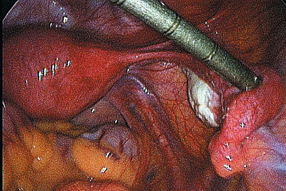
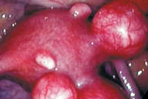
Endometriosis
Laparoscopic Image of Endometriosis
Note brownish-black “powder burn” implants. (Courtesy of Dr. S. Kives and Dr. R. Spitzer)
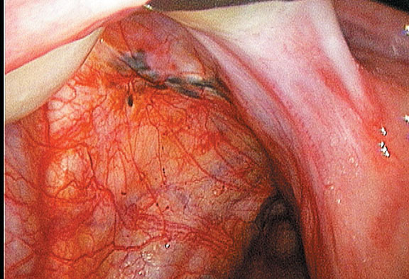
Leiomyoma – Microscopic
Microscopic view of proliferative smooth muscle cells. (Courtesy of Dr. I. Zberiranowski)
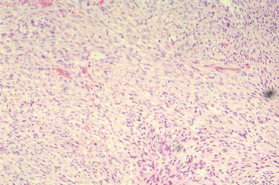
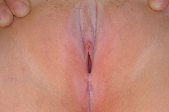
Lichen Sclerosis
Note classic hour glass or figure 8 vulvular and perianal distribution. (Courtesy of Dr. S. Kives and Dr. R. Spitzer)
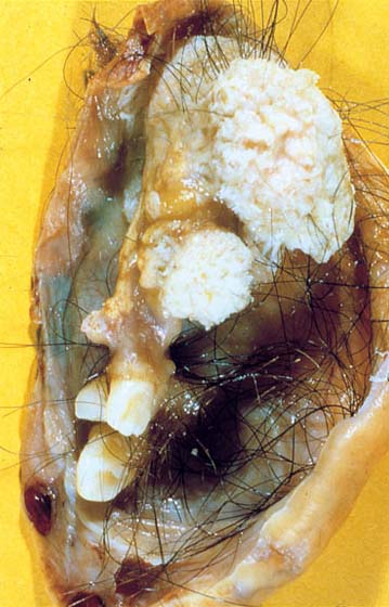
Ovarian Teratoma
Cystic Teratoma
Gross appearance of an ovary with a mature cystic teratoma. (Courtesy of Dr. I. Zberiranowski)
