Browse by specialty
- Anesthesia and Perioperative
- Cardiology & Cardiovascular System
- Clinical Pharmacology
- Dermatology
- Diagnostic Imaging
- Emergency Medicine
- Endocrinology
- Ethical, Legal, and Organizational Medicine
- Family Medicine
- Gastroenterology
- General Surgery
- Geriatric Medicine
- Gynecology
- Haematology
- Infectious Disease
- Nephrology
- Neurology
- Neurosurgery
- Obstetrics
- Ophthalmology
- Orthopedics
- Otolaryngology
- Pediatrics
- Plastic Surgery
- Population and Community Health
- Psychiatry
- Respirology
- Rheumatology
- Urology
Gastroenterology
Full-Colour Atlas
Mouth
Aphthous ulcer of Crohn’s disease. Note normal surrounding mucosa.
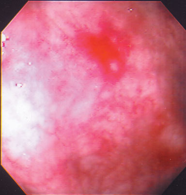
Photo courtesy of Dr. G. Kandel.
Esophagus
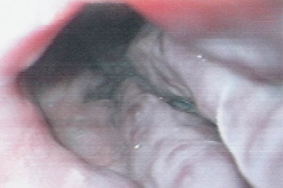
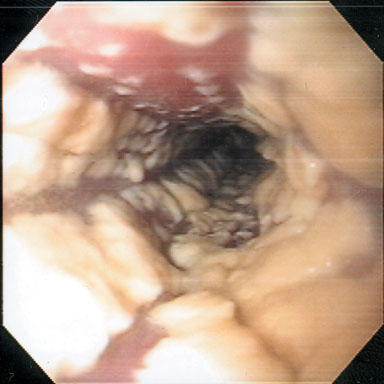
| Esophageal Varices | Candida Esophagitis |
|---|---|
 |
 |
| Barrett's Esophagus | |
| Columnar epithelium extends up into normal squamous epithelium in one quadrant.
|
All photos courtesy of Dr. G. Kandel.
Stomach
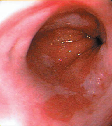
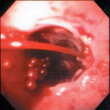
| Peptic Ulcer Disease | Bleeding Ulcer |
|---|---|
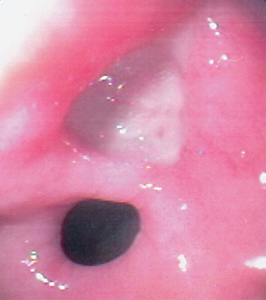 |
Blood spurting from a small ulcer.
|
All photos courtesy of Dr. G. Kandel.
Liver Disease
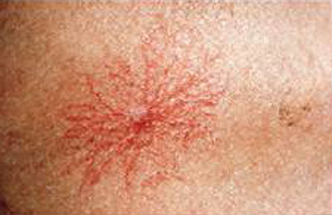
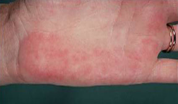
| Spider Nevi | Palmar Erythema |
|---|---|
 |
 |
| Leukonychia | Ascites |
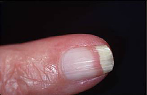 |
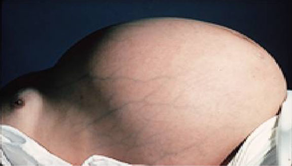 |
| Caput Medusae | Clubbing |
| Please see image at: https://www.healthline.com/health/caput-medusae | Please see image at: https://en.wikipedia.org/wiki/Nail_clubbing |
| Dupuytren’s Contracture | Scleral Icterus |
| Please see image at: https://floridahandcenter.com/dupuytrens-contracture/ | Please see image at: https://www.thesilverfridge.com/medical-trivial-repository/2020/1/3/gilberts |
| Parotid Enlargement | Liver Cirrhosis |
| Please see image at: https://www.sciencephoto.com/media/c0111729/view | Please see image at: https://my.clevelandclinic.org/-/scassets/images/org/health/articles/15572-cirrhosis?io=transform%3afit%2cwidth%3a780 |
Colon
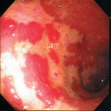
| Pseudomembranous Colitis | Ulcerative Colitis |
|---|---|
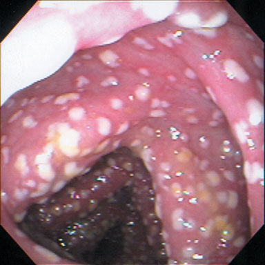 |
Diffuse, erythema, friability and loss of normal vascular pattern.
|
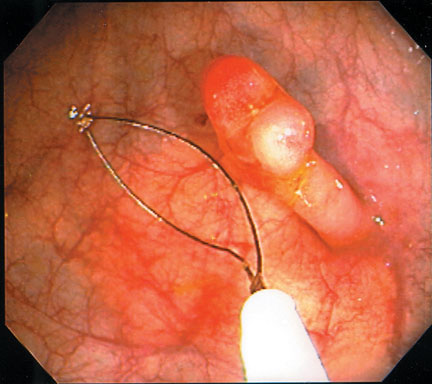
| Colonic Polyp | Angiodysplasia (“Watermelon Stomach”) |
| Removal with snare.
|
Usually presents as anemia and can be treated by endoscopic coagulation
|
All photos courtesy of Dr. G. Kandel.
Rectum

