Browse by specialty
- Anesthesia and Perioperative
- Cardiology & Cardiovascular System
- Clinical Pharmacology
- Dermatology
- Diagnostic Imaging
- Emergency Medicine
- Endocrinology
- Ethical, Legal, and Organizational Medicine
- Family Medicine
- Gastroenterology
- General Surgery
- Geriatric Medicine
- Gynecology
- Haematology
- Infectious Disease
- Nephrology
- Neurology
- Neurosurgery
- Obstetrics
- Ophthalmology
- Orthopedics
- Otolaryngology
- Pediatrics
- Plastic Surgery
- Population and Community Health
- Psychiatry
- Respirology
- Rheumatology
- Urology
Dermatology
Full-Colour Atlas
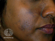
Acne Vulgaris
Inflammatory papules, pustules, and open comedones.
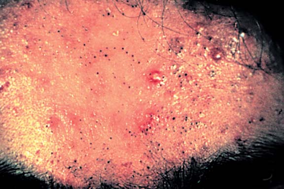
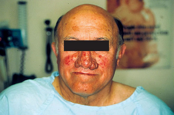
Rosacea
Prominent facial erythema, telangiectasia, rhinophyma, and scattered papules. (Courtesy Dr. L. From)
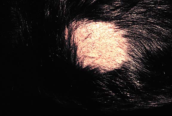
Tinea Capitis
Diffuse area of mild scaling and hair loss. Erythema and pyoderma are occasionally present.
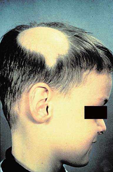
Alopecia Areata
Sharply demarcated, circular patch of hair loss on scalp. Look for “exclamation mark” hairs at the periphery.
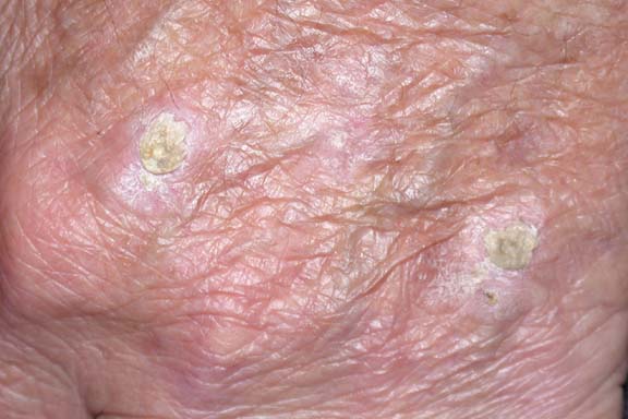
Actinic Keratosis – Hand
Hyperkeratotic, erythematous, slightly elevated papules and patches with a rough surface on sun-exposed skin. (Courtesy Dr. S. Walsh)
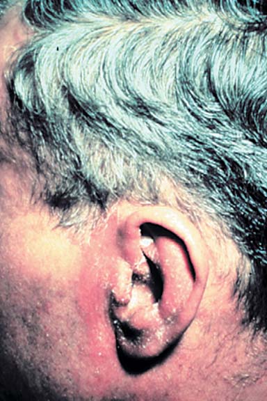
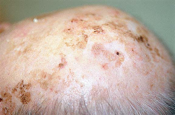
Actinic Keratosis – Head
Hyperkeratotic, erythematous, slightly elevated papules and patches with a rough surface on sun-exposed skin. (Courtesy Dr. C. Forrest)
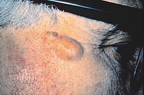
Epidermal Cyst
Round, firm, yellow/flesh coloured, mobile nodule; may observe a follicular punctum on the overlying epidermal surface.
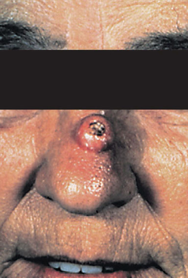
Keratoacanthoma
Erythematous, skin-coloured, firm, dome-shaped nodule with central keratin-filled crater.
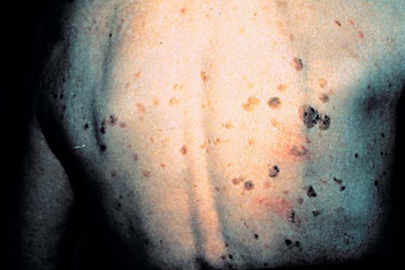
Seborrheic Keratosis
Well-demarcated, waxy, brownish-black or tan papules/plaques; warty and “stuck-on” appearance.
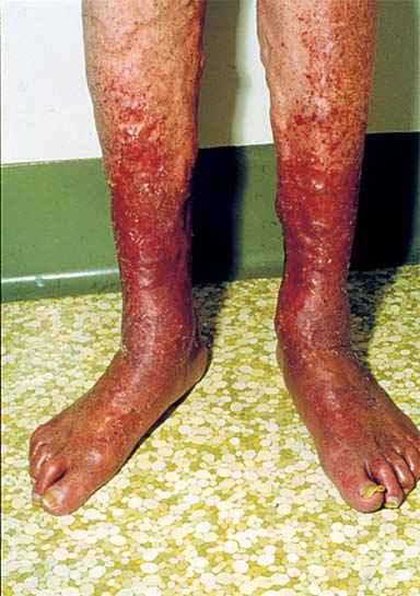
Stasis Dermatitis
Erythematous scaling patches. May see with hyperpigmentation, swelling, and ulceration. (Courtesy of Dr. L. From)
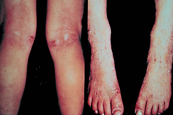
Atopic Dermatitis
Excoriated, lichenified plaques with erythema, dryness, and crusting.
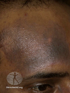
Hyperpigmentation due to atopic dermatitis (eczema).
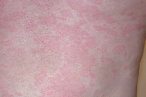
Urticaria
Circumscribed, raised, edematous, red plaques surrounded by faint white halo. (Courtesy Dr. S. Walsh)
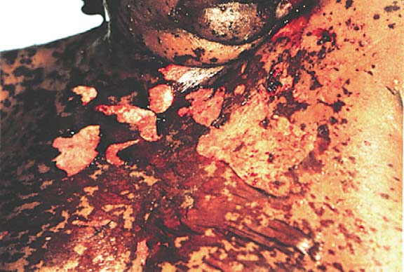
Toxic Epidermal Necrolysis – TEN
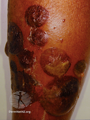
Widespread necrosis with painful blistering and denuding of epidermis.
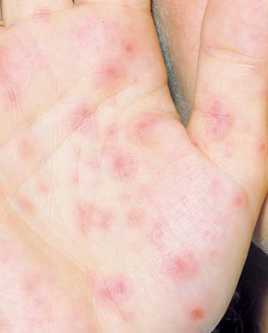
Erythema Multiforme EM No.2
The macules and papules can have various appearances but tend to be monomorphic in a given patient. (Courtesy Dr. S. Walsh)
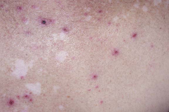
Erythema Multiforme EM – No.1
Macules/papules with concentric, light and dark erythematous rings. (Courtesy of Women’s College Hospital Slide Library, Toronto)
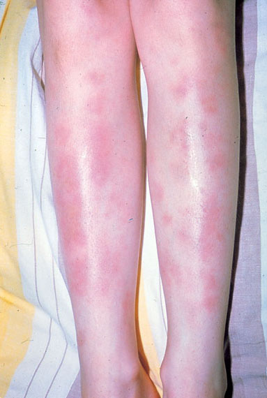
Erythema Nodosum
Tender, poorly demarcated, deep-seated nodules usually on lower extremities. (Courtesy Dr. M. Mian)
Heritable Disorders
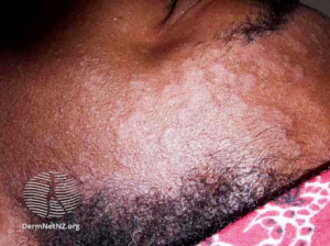
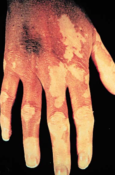
Vitiligo
Sharply marginated, white patches completely devoid of pigment due to an acquired loss of melanocytes.
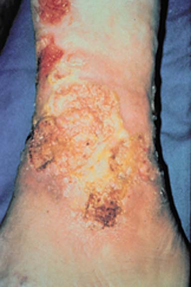
Squamous Cell Carcinoma
Indurated erythematous nodule or plaque with a hyperkeratotic surface, scale/crust, and central ulceration.

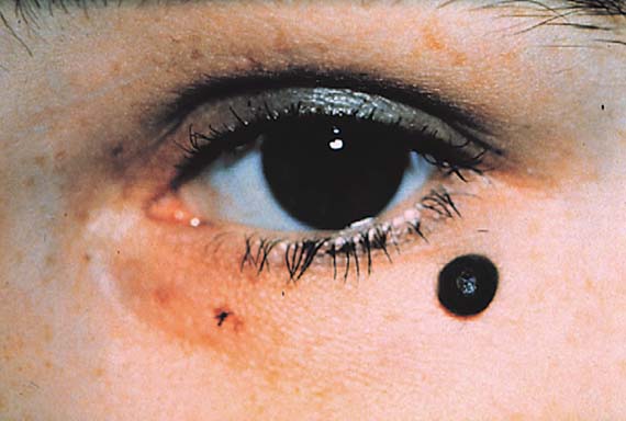
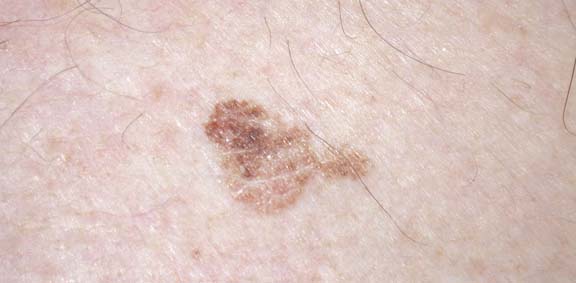
Malignant Melanoma
A pigmented lesion with asymmetry, an irregular border, variegated colour, and diameter greater than 6 mm.
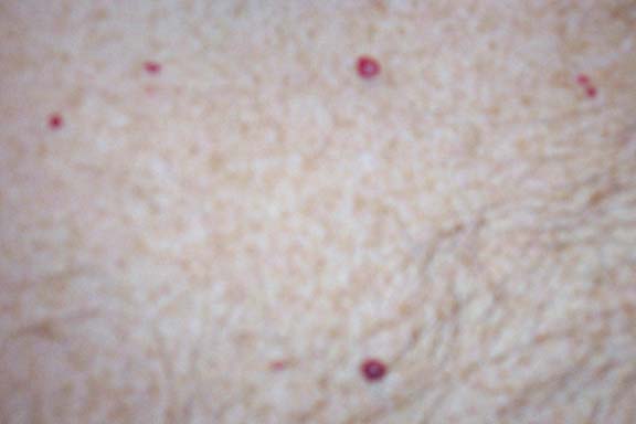
Basal Cell Carcinoma No. 2
Skin-coloured papule or plaque with rolled, translucent/pearly border and telangiectasia.
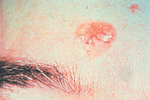
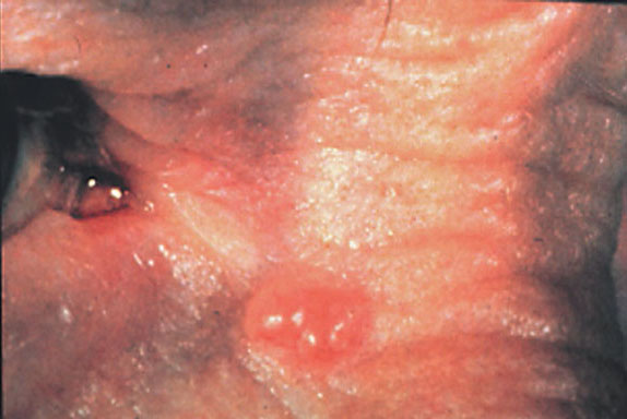
Basal Cell Carcinoma No. 1
Skin-coloured papule or plaque with rolled, translucent/pearly border and telangiectasia.
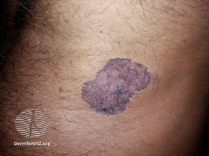
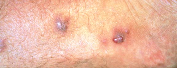
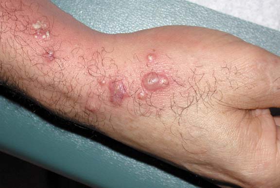
Pustular Psoriasis
Deep-seated, dusky-red macules and creamy-yellow pustules progress to hyperkeratotic/crusted papules. (Courtesy Dr. S. Walsh)
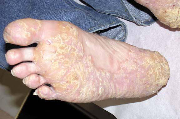
Psoriasis of the Soles
Well-demarcated, erythematous plaques with thick, yellowish scale and desquamation on sites of pressure arising on the plantar surface of feet. (Courtesy Dr. S. Walsh)

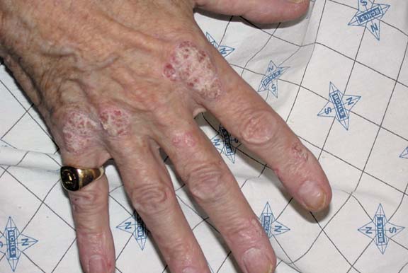
Plaque Psoriasis – Hand
Dry, well-circumscribed, silvery scaling, erythematous papules and plaques. (Courtesy Dr. S. Walsh)
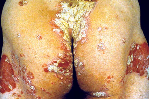
Plaque Psoriasis – Body
Dry, well-circumscribed, silvery scaling, erythematous papules and plaques. (Courtesy Dr. L. From)

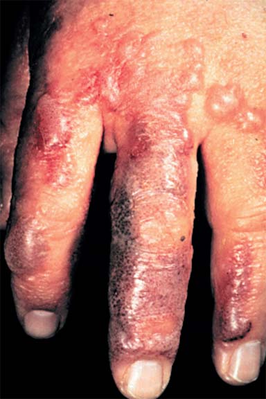
Lichen Planus
Violaceous, flat-topped polygonal papules on the flexural surface of the wrists. (Courtesy Dr. S. Walsh)
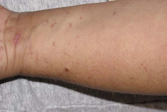
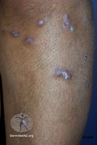
Pemphigus Vulgaris
Flaccid vesicles and bullae that easily rupture; erosions and crusts. (Courtesy Dr. S. Walsh)
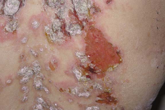

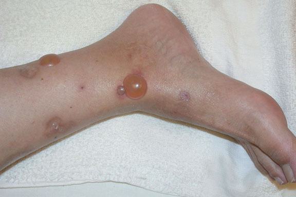
Bullous Pemphigoid
Multiple tense serous and partially hemorrhagic bullae; post-inflammatory hyperpigmentation is present at sites of prior lesions. Note the presence of uninvolved skin between lesions. (Courtesy Dr. S. Walsh)
