Browse by specialty
- Anesthesia and Perioperative
- Cardiology & Cardiovascular System
- Clinical Pharmacology
- Dermatology
- Diagnostic Imaging
- Emergency Medicine
- Endocrinology
- Ethical, Legal, and Organizational Medicine
- Family Medicine
- Gastroenterology
- General Surgery
- Geriatric Medicine
- Gynecology
- Haematology
- Infectious Disease
- Nephrology
- Neurology
- Neurosurgery
- Obstetrics
- Ophthalmology
- Orthopedics
- Otolaryngology
- Pediatrics
- Plastic Surgery
- Population and Community Health
- Psychiatry
- Respirology
- Rheumatology
- Urology
Cardiology & Cardiovascular System
Full-Colour Atlas
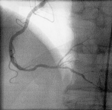
Normal RCA
Normal RCA with posterior interventricular (PIV) artery running inferiorly, and the posterior lateral branches branching superiorly in this view. (Courtesy of Toronto General Hospital Catheterization Laboratory)
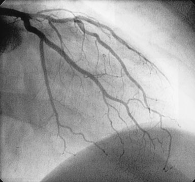
Normal LCA
Normal left coronary artery (LCA) with the circumflex system inferior to the left anterior descending (LAD) artery in this view. (Courtesy of Toronto General Hospital Catheterization
Normal RCA
Normal RCA with posterior interventricular (PIV) artery running inferiorly, and the posterior lateral branches branching superiorly in this view. (Courtesy of Toronto General Hospital Catheterization Laboratory)

Normal LCA
Normal left coronary artery (LCA) with the circumflex system inferior to the left anterior descending (LAD) artery in this view. (Courtesy of Toronto General Hospital Catheterization

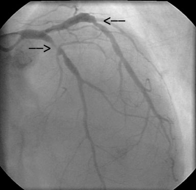
Stents Installed
This patient had two stents installed in this LAD, which you can see as cylindrical shapes within the vessel.
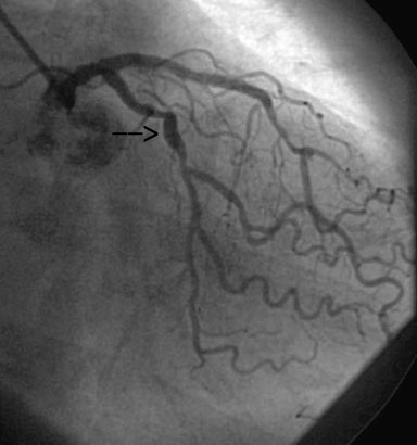
Stenotic Lesion
LAD with 95% stenotic lesion distal to diagonal branches. (Courtesy of Toronto General Hospital Catheterization Laboratory)
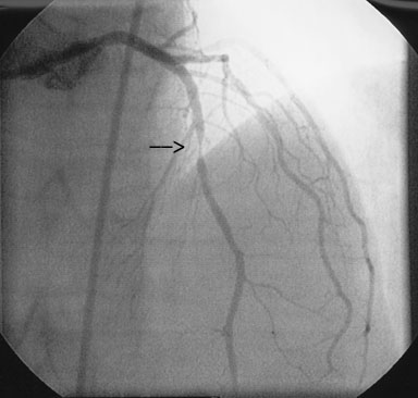
Stenosis LAD
Stenosis of LAD, as well as the circumflex.
Echocardiography
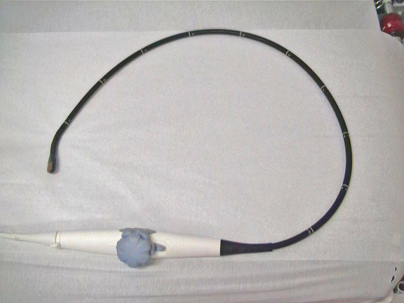
TEE Probe
Probe used in transesophageal echocardiogram. (Courtesy of Dr. Chi-Ming Chow)
Echocardiogram Video
TTE: Transthoracic echocardiogram with Doppler ultrasound showing the four chambers and blood flow. (Courtesy of Dr. Chi-Ming Chow)
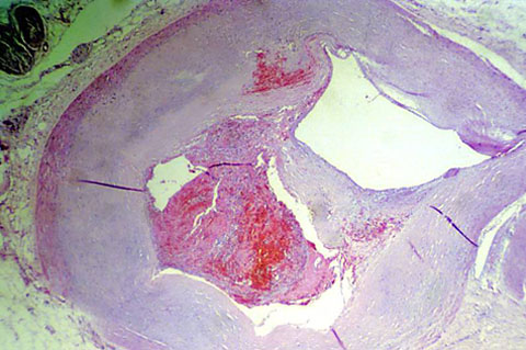
Atherosclerosis Histology
Histological view of an atherosclerotic fatty plaque. (courtesy of Dr. Jagdish Butany)
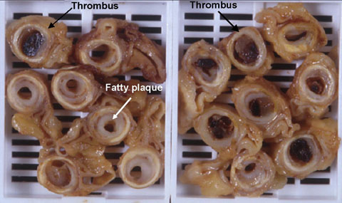
Atherosclerosis Cross Sections
Cross-sections of multiple arteries from a patient with atherosclerosis, seen as white fatty plaque occluding the arterial lumen. Various degrees of stenosis are seen. Some of vessels showed evidence of plaque rupture and formation of a fully occluding thrombus (black arrows). (courtesy of Dr. Jagdish Butany)
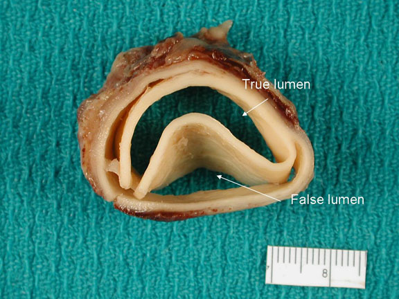
Aortic Dissection
Gross anatomical view of an aortic dissection. The true and false lumens of the dissected aorta are shown (courtesy of Dr. Jagdish Butany)
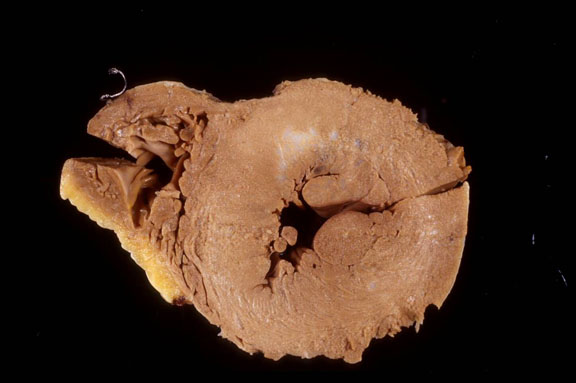
Hypertrophic Cardiomyopathy
The ventricular wall is much thicker compared to that of a normal heart. (courtesy of Dr. Jagdish Butany)
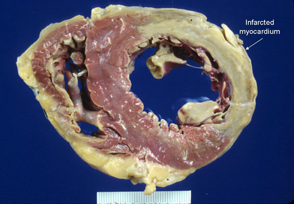
Dilated Cardiomyopathy
The ventricle is enlarged with normal thickness of the ventricular wall. Note that this gross specimen also shows a lateral wall infarct stained as white myocardial tissue.
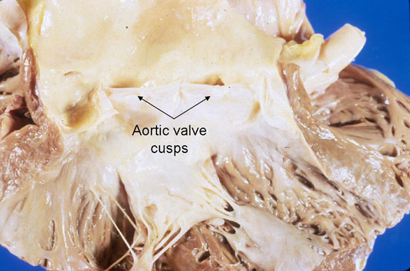
Normal aortic valves
(courtesy of Dr. Jagdish Butany)
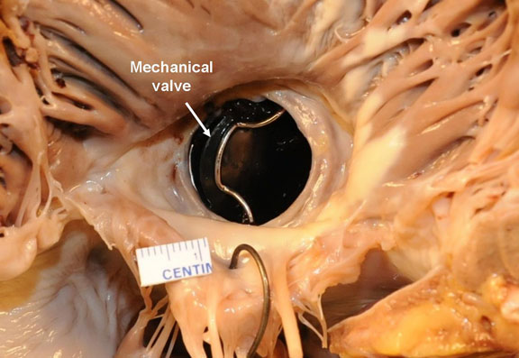
Mechanical Valve In Situ
In this specimen, the aortic valves were replaced with the metallic (Bjork-Shiley) valve. (courtesy of Dr. Jagdish Butany)
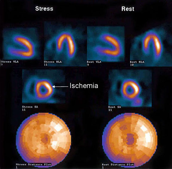
Perfusion Scan
Perfusion scan showing blood flow during rest and persantine stress. White areas mark those of ischemia. Irreversible defects are seen with ischemia both at rest and stress. (courtesy of Dr. Chi-Ming Chow)
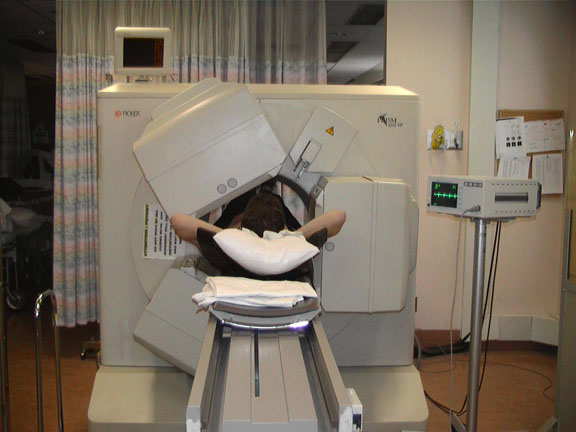
Cardiac Nuclear Scan
Patient undergoing single photon emission tomography for nuclear cardiac imaging. (courtesy of Dr. Chi-Ming Chow)
ECG Made Simple (EMS) is a comprehensive web-based ECG learning program that teaches the art and science of electrocardiogram (ECG) interpretation.
iCCS App
The iCCS app for iOS and Android includes Guidelines and KT resources for dyslipidemia, heart failure, atrial fibrillation, and peripheral arterial disease.
