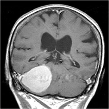
T1-weighted coronal MRI with contrast. A large tumour is highlighted in the right posterior fossa and bilateral dilatation of the lateral ventricles is evident (likely secondary to compression of the 4th ventricle or Sylvian aqueduct). [Courtesy of Dr. J. Spears]
Coloured Atlas
- Cardiology & CVS
- Coloured Atlas – Dermatology
- Coloured Atlas – Endocrinology
- Gastroenterology (Coloured Atlas)
- Geriatric Medicine
- Coloured Atlas – Gynecology
- Coloured Atlas – Hematology
- Coloured Atlas – Infectious Disease
- Coloured Atlas – Nephrology
- Neurology
- Coloured Atlas – Neurosurgery
- Coloured Atlas – Obstetrics
- Coloured Atlas – Ophthalmology
- Coloured Atlas – Orthopedics
- Coloured Atlas – Otolaryngology
- Coloured Atlas – Pediatrics
- Coloured Atlas – Plastic Surgery
- Coloured Atlas – Rheumatology
- Urology