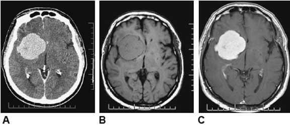
Well-demarcated, homogeneous, extra-axial lesion.
a) CT
b) T1 weighted MRI (note that the tumour is isointense to the gray matter)
c) T1 weighted MRI post contrast.
[Courtesy of Dr. W. Montanera]

by Tim Milligan | Nov 13, 2015 | Tumours
Well-demarcated, homogeneous, extra-axial lesion.
a) CT
b) T1 weighted MRI (note that the tumour is isointense to the gray matter)
c) T1 weighted MRI post contrast.
[Courtesy of Dr. W. Montanera]
