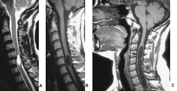
Image A: T2 weighted MRI
Image B: T1 weighted MRI
Image C: T1 weighted MRI of another case with upper arrow indicating Chiari malformation. [Courtesy of Dr. W. Montanera]

by Tim Milligan | Nov 13, 2015 | Spine
Image A: T2 weighted MRI
Image B: T1 weighted MRI
Image C: T1 weighted MRI of another case with upper arrow indicating Chiari malformation. [Courtesy of Dr. W. Montanera]
