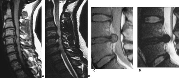
Image A: T1 and
Image B: T2 weighted MRI of degenerative cervical disc herniation
Image C: Intermediate and
Image D: T2 weighted MRI demonstrating large disc herniation with spinal cord compression.
[Courtesy of Dr. W. Montanera]

by Tim Milligan | Nov 13, 2015 | Spine
Image A: T1 and
Image B: T2 weighted MRI of degenerative cervical disc herniation
Image C: Intermediate and
Image D: T2 weighted MRI demonstrating large disc herniation with spinal cord compression.
[Courtesy of Dr. W. Montanera]
