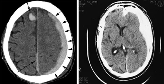
Non-contrast axial CT (left image) showing a left hemispheric subdural hematoma with some blood tracking along the falx and concurrent intracerebral hemorrhage on the right.
Some compression is seen on the left with decreased visibility of sulci and soft tissue swelling on the posterior left occipital lobe.
The right image shows left increased density, concave mass usually less uniform, less dense, and more diffuse than epidural hemorrhage. Note compression of ventricles and midline shift. [Courtesy of Dr. J. Spears]