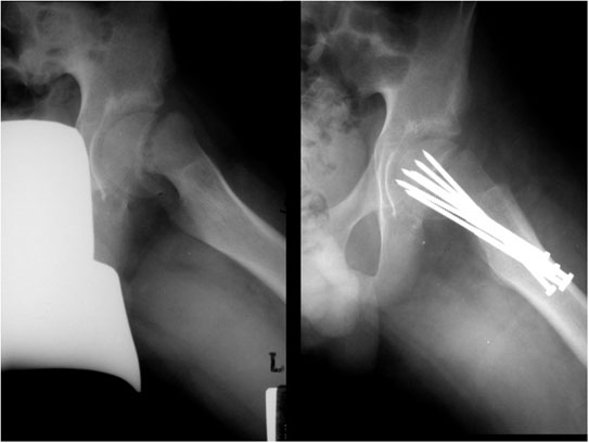
The image to the left is of a slipped capital femoral epiphysis in a child. The image to the right is after fixation. [Courtesy of Dr. Tim Dowdell, St. Michael’s Hospital, Department of Medical Imaging]

by Tim Milligan | Nov 12, 2015 | Traumatic Lower Extremity
The image to the left is of a slipped capital femoral epiphysis in a child. The image to the right is after fixation. [Courtesy of Dr. Tim Dowdell, St. Michael’s Hospital, Department of Medical Imaging]
