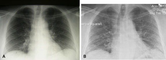
The left image (a) shows mildly reduced lung volumes, hazy opacifications and reticulation, primarily in the lower lobes. The right image (b), taken about 9 months later, shows progression of the reticulation, volume loss and some nodularity. [Courtesy of Dr. Ted Marras]

