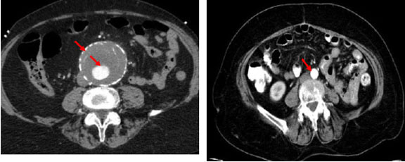
Left Image: Abdominal axial CT with contrast demonstrating a large AAA with extensive intraluminal thrombosis. Patent central lumen containing contrast surrounded by thrombus peripherally. Aortic wall evident due to circumferential calcification (atherosclerosis).
Right Image: Abdominal axial CT with contrast showing abdominal aorta of normal diameter.
[Courtesy of Dr. N. Jaffer]

