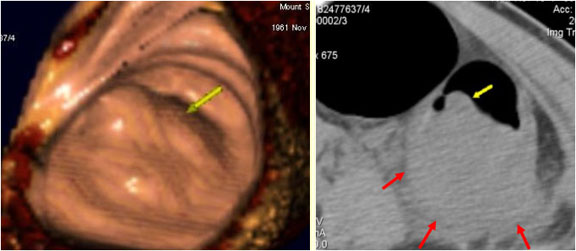
CT colonography (virtual colonoscopy). 3-D endoluminal and 2-D axial CT images shown in parallel for same colonic lesion. Images demonstrate the importance of comparing both images for determining extent of identified lesions. Endoluminal view shows intraluminal part of lesion only, while the axial image demonstrates serosal extension of the lesion allowing for more accurate staging (arrows). [Courtesy of Dr. N. Jaffer]
Coloured Atlas
- Cardiology & CVS
- Coloured Atlas – Dermatology
- Coloured Atlas – Endocrinology
- Gastroenterology (Coloured Atlas)
- Geriatric Medicine
- Coloured Atlas – Gynecology
- Coloured Atlas – Hematology
- Coloured Atlas – Infectious Disease
- Coloured Atlas – Nephrology
- Neurology
- Coloured Atlas – Neurosurgery
- Coloured Atlas – Obstetrics
- Coloured Atlas – Ophthalmology
- Coloured Atlas – Orthopedics
- Coloured Atlas – Otolaryngology
- Coloured Atlas – Pediatrics
- Coloured Atlas – Plastic Surgery
- Coloured Atlas – Rheumatology
- Urology