A. Peripheral visual field
(a) wiggling fingers
(b) counting fingers
(c) white pin
B. Central visual field
(a) red pin
Examination Technique:
- visual fields are assessed by confrontation , i.e. the examiner compares the patient’s visual field to their own and assumes that theirs is normal.
- first test each eye separately.
- test both eyes together with wiggling fingers.
- the examiner places himself approximately 1 meter away from the patient and advises the patient to look directly at the examiner’s eye for monocular testing or nose for binocular testing. The test object (either a wiggling finger, one or two fingers, or a white pin head) is presented equidistant from the patient’s and examiner’s eye and the patient is asked either to state the number of fingers or say “yes” when they first see a moving target.
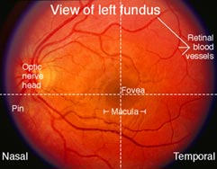
- for central vision (the 20 degrees on either side of the vertical meridian) a red pin is used. The patient is instructed to state when they see the pin as red. A red pin is also used to map the blind spot. Vision in the center of the visual field is more detailed than in the peripheral areas. This is because of both the structure of the retina and the connections of its neurons. Light rays from the center of the visual field are focused on the macula in the center of the retina. In the macula, the proportion of cones to rods is high. Cones are important for color vision.
Normal Response:
the normal peripheral monocular visual field extends approximately 90 degrees temporally, 60 degrees nasally, 60 degrees superiorly and 75 degrees inferiorly. It is divided into nasal and temporal halves and superior and inferior altitudinal halves. The normal central visual field extends approximately 30 degrees on all sides of central fixation. The blind spot is located 15 degrees temporal to fixation just below the horizontal meridian. It corresponds to the optic disc.