As pulmonary edema fluid initially collects in the interstitium, mild pulmonary edema shows the following features:
- loss of definition of pulmonary vasculature
- peribronchial cuffing
- Kerley B lines
- reticulonodular pattern
- thickening of interlobar fissures
If the pulmonary edema is due to heart failure or fluid overload, you may also see cardiomegaly and distension of the pulmonary veins, particularly in the upper lung fields. This latter phenomenon is referred to as vascular redistribution and can be seen in normal supine radiographs of the chest. As pulmonary edema progresses, fluid begins to collect in alveoli, causing diffuse airspace disease often in a “bat-wing” or “butterfly” pattern, tending to spare the outermost lung fields. Pleural effusion may be a feature of pulmonary edema.
Left: Normal
Right: Enlarged upper lobe vessels
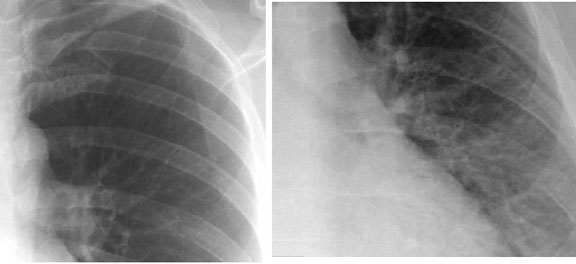
Pulmonary Vasculature Redistribution