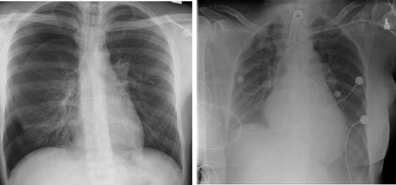
The patient’s left pneumothorax is more difficulty to diagnose on this supine film. This study demonstrates the “deep sulcus” sign, with the left costophrenic sulcus descending below the edge of the film. Other clues include a hyperlucent left hemithorax and slight sharpening of the left mediastinal border. This patient also has a tracheostomy, evidence of a prior sternotomy (not the multiple circular sternal wires), and a central venous catheter with its tip in the right ventricular outflow tract. The circular structures projecting over the chest, some with wires extending from them are ECG leads.
Pneumothorax – Pleural Disease