In a normal chest x-ray, the diaphragm and mediastinal structures are visible because of the difference in radiodensity between lung and these structures (i.e. there is an “interface” between the tissues). The “silhouette” sign refers to loss of normally appearing interfaces, implying opacification due to consolidation (most common), atelectasis, mass, etc., in adjacent lung.
This patient demonstrates “silhouetting” of the right heart border to right middle lobe consolidation. Note that the right hemidiaphragm is still well seen.
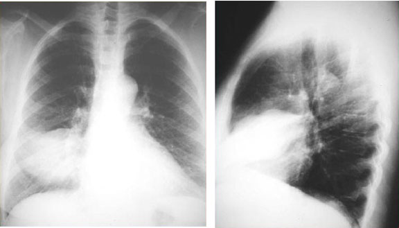
Silhouette Sign – RML