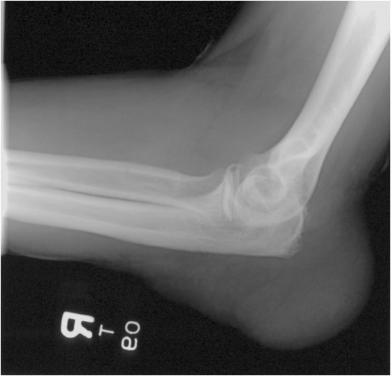
Radiology

Figure – 11

Elbow radiograph in a patient with gout. Note large soft tissue abnormality (arrows) superficial to the olecranon. This represents olecranon bursitis. In addition to gout, it can also be seen in patients with rheumatoid arthritis, or be related to trauma or infection. Bilateral olecranon bursitis is characteristic for gout. (Courtesy of Dr. A. Donovan)

Elbow radiograph

Figure – 10

Left toe radiograph shows well-marginated erosions with sclerotic margins (arrow) and overhanging edges (arrowheads) at the interphalangeal joint. The findings are consistent with gouty arthritis. Similar changes are seen at the first MTP joint. (Courtesy of Dr. A. Donovan)

Left toe radiograph

Figure – 1

Bilateral hand radiographs in a patient with osteoarthritis. There is moderate joint space loss at the first carpometacarpal (CMC) joints bilaterally (arrows) with subchondral sclerosis and cystic changes. The interphalangeal joints show similar changes. These are shown by the arrowhead. (Courtesy of Dr. A. Donovan)

Bilateral hand radiographs