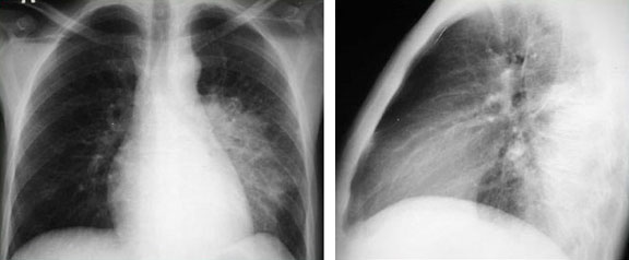
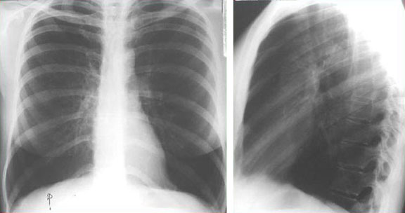
Airspace

Spine Sign

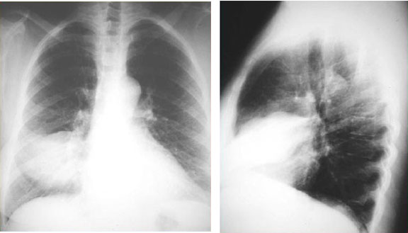
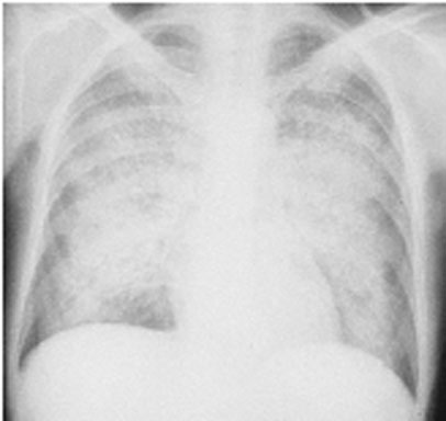
This young patient has left lower lobe pneumonia. The left hemidiaphragm is “silhouetted” by consolidation in the left lower lobe (note that one cannot see the entire left hemidiaphragm through the cardiac shadow).
In a normal chest x-ray, the diaphragm and mediastinal structures are visible because of the difference in radiodensity between lung and these structures (i.e. there is an “interface” between the tissues).
The “silhouette” sign refers to loss of normally appearing interfaces, implying opacification due to consolidation (most common), atelectasis, mass, etc., in adjacent lung.

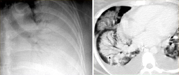
The lateral film demonstrates the “spine” sign. On a normal lateral chest x-ray, as one moves down the thoracic vertebral column, the vertebral bodies appear progressively blacker. Here they appear more radioopaque due to consolidation in the overlying left lower lobe.

Spine Sign

Silhouette Sign – RML

In a normal chest x-ray, the diaphragm and mediastinal structures are visible because of the difference in radiodensity between lung and these structures (i.e. there is an “interface” between the tissues). The “silhouette” sign refers to loss of normally appearing interfaces, implying opacification due to consolidation (most common), atelectasis, mass, etc., in adjacent lung.

This patient demonstrates “silhouetting” of the right heart border to right middle lobe consolidation. Note that the right hemidiaphragm is still well seen.

Silhouette Sign - RML

Pulmonary Edema

The plain AP films of this patient exemplify pulmonary edema. One can easily appreciate the fluffy white opacities throughout the lung field. Other signs such as vascular redistribution, peri-bronchial cuffing, and pleural effusion are difficult to appreciate on this study. However, Kerley B lines are seen, especially in the lower right lung field.

Pulmonary Edema

Hyperinflation

This young patient has emphysema as a result of alpha-1-antitrypsin deficiency. Hyperinflation is noted as a generalized increase in radiolucency due to increased aeration and spread of vasculature, an increased AP chest diameter and retrosternal airspace on the lateral view, and diaphragmatic domes projecting well below the normal level of the 10th rib posteriorly and the 6th rib anteriorly on the PA view.

Hyperinflation

Bullae

Bullae are often associated with emphysema. By definition, a bulla is a gas-containing, avascular area of lung at least 1 cm in diameter and with a wall thickness of at least 1 mm. The arrows indicate bullae visible on the chest x-ray (left) and CT scan (right) of a patient with bullous emphysema.

Bullae