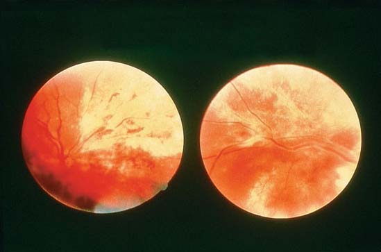
Retinal Detachment
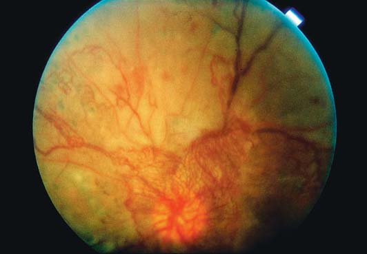
Bullous retinal detachment with retinal folds on temporal aspect.
Study Smarter
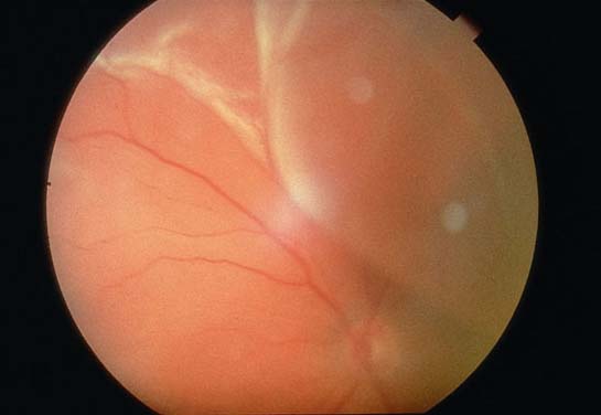
Bullous retinal detachment with retinal folds on temporal aspect.

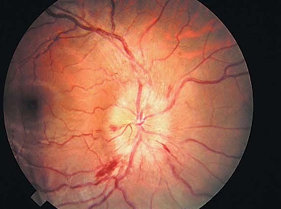
Elevated congested disc with indistinct margins, flameshaped hemorrhages, and dilated tortuous vessels.
Pallor of optic disc with sharp margin; attenuated vessels.
(NVD) Early fluorescein angiography image of the eye in image OP35 demonstrating areas of vascular leakage near the disc. 1. NVD; 2. Vascular leakage
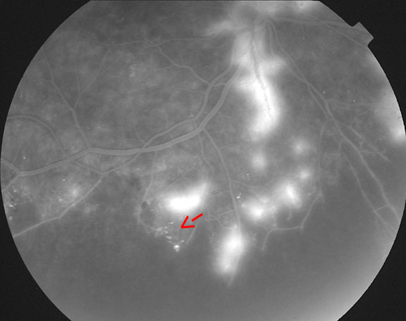
(NVE) Fluorescein angiography image of the eye in image OP35 showing areas of vascular leakage.
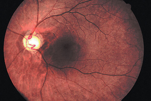
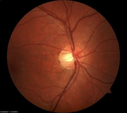
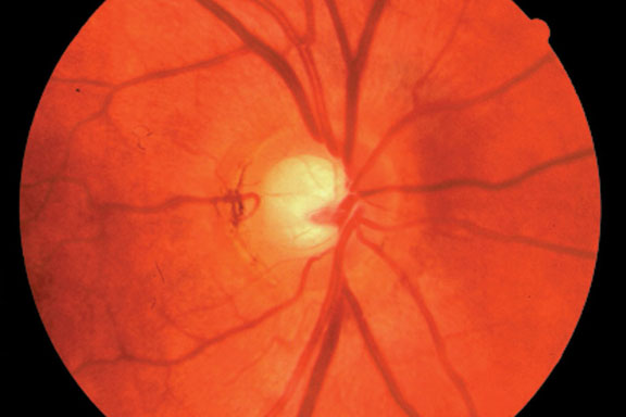
Asymmetrical increase of cup: disc ratio (0.8). Cupping seen where vessels disappear over the edge of the attenuated rim.
Proliferative Diabetic Retinopathy
Fan-shaped network of new blood vessels branching onto optic disc (neovascularization). Also note dot hemorrhages and microaneurysms.
Proliferative Diabetic Retinopathy
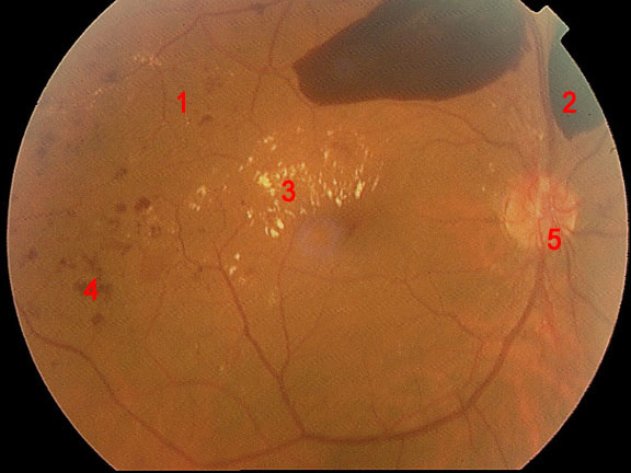
Proliferative Diabetic Retinopathy
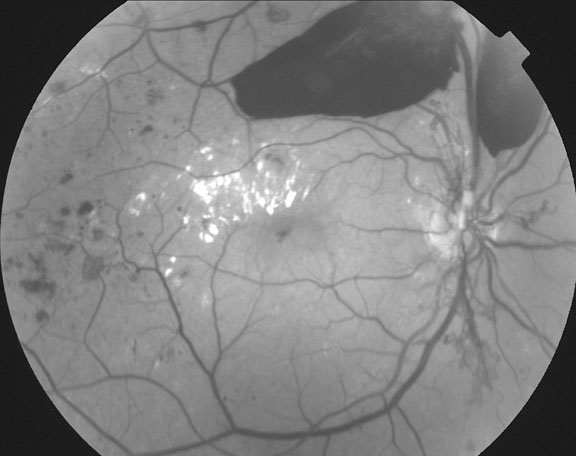
Red-free image of previous image Proliferative Diabaetic Retinopathy.
White exudates surrounding hemorrhages and areas of necrosis. Distinct border between diseased and normal retina.
