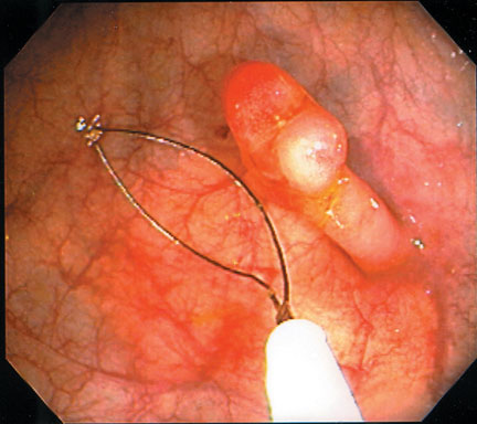
Esophageal Varices
Esophageal Varices (Courtesy of Dr. G. Kandel)
Study Smarter
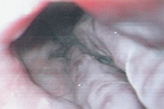
Esophageal Varices (Courtesy of Dr. G. Kandel)

(Courtesy of Dr. G. Kandel)
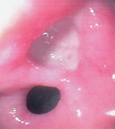
Blood spurting from a small ulcer. (Courtesy of Dr. G. Kandel)
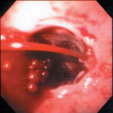
Apthous ulcer of Crohn’s disease. Note: normal surrounding mucosa. (Courtesy of Dr. G. Kandel)
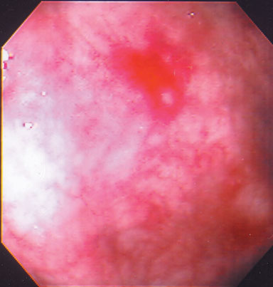
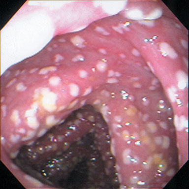
Pseudomembranous Colitis (Courtesy of Dr. G. Kandel)
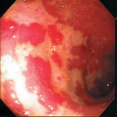
Ulcerative Colitis
Diffuse, erythema, friability and loss of normal vascular pattern. (Courtesy of Dr. G. Kandel)
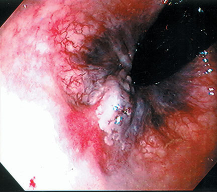
Internal Hemorrhoid
View by retroflexing colonscope. (Courtesy of Dr. G. Kandel)
(Courtesy of Dr. G. Kandel)
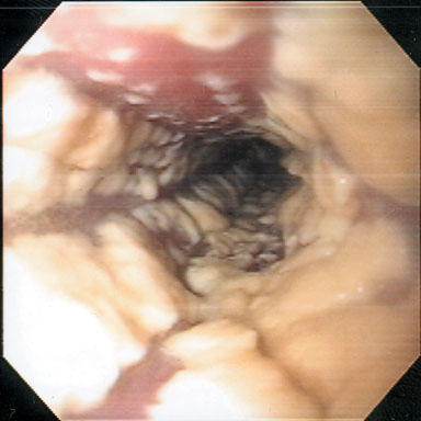
Columnar epithelium extends up into normal squamous epithelium in one quadrant. (Courtesy of Dr. G. Kandel)
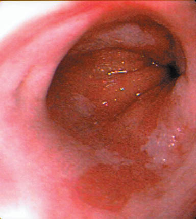
Removal with snare. (Courtesy of Dr. G. Kandel)
