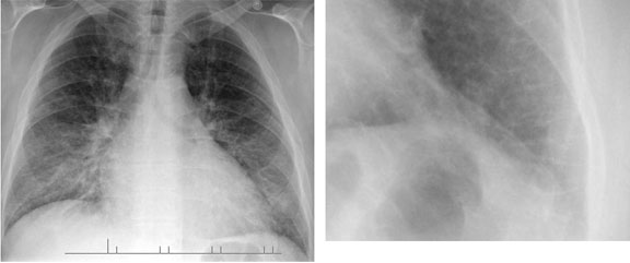
Lung Parenchyma

Pulmonary Nodules

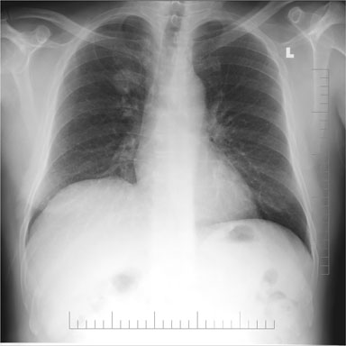
This image shows a pulmonary nodule in the right upper lung. Differential diagnosis of pulmonary nodules includes:

  1. Malignancy (primary or metastatic)
  2. Benign neoplasm: hamartoma, bronchial adenoma
  3. Granuloma
  4. Simulated: nipple, bone lesion, skin lesion, foreign body, artefact

Less common causes include:

  1. Abscess
  2. Infarct
  3. Loculated pleural effusion
  4. Organized pneumonia
  5. Sarcoidosis
  6. Cystis disease
  7. Vascular lesions

Pulmonary Nodules

Pulmonary Edema – Kerly B Lines

Kerly lines result fomr thickened connective tissue planes occurring as a result of pulmonary edema or carcinomatosis.

Kerly A Lines: Radiate towards hila in mid- and upper-lung zones, 3-4 cm long, smaller than vascular markings (not useful)
Kerly B Lines: Horizontal, <2 cm long an d1 mm thick, at periphery of lung, reach lung edge (very useful)
Kerly C Lines: Thickened interlobular septa

Pulmonary Edema - Kerly B Lines

Interstitial Disease – Kerly B Lines

Kerly lines result fomr thickened connective tissue planes occurring as a result of pulmonary edema or carcinomatosis.

Kerly A Lines: Radiate towards hila in mid- and upper-lung zones, 3-4 cm long, smaller than vascular markings (not useful)
Kerly B Lines: Horizontal, <2 cm long an d1 mm thick, at periphery of lung, reach lung edge (very useful)
Kerly C Lines: Thickened interlobular septa

Interstitial Disease - Kerly B Lines

Interstitial Disease

Interstitial disease is a pathological process involving the interlobular connective tissue (i.e. scaffolding of the lung).

Differential diagnosis of interstitial disease includes:

  1. Pulmonary edema (which can move into the alveoli)
  2. Collagen disease (fibrosis)
  3. Sarcoidosis
  4. Pneumoconiosis
  5. Metastatic disease (from the lymphatics)
  6. Inflammatory conditions (viral/interstitial pneumonia)

Interstitial Disease