Neurological Exam

Tone

Examination Technique:

  • ensure the patient is relaxed.
  • for assessment in the upper extremities, the patient may be lying or sitting. In the lower extremities, tone is best assessed with the patient lying down.
  • explain the examination technique to the patient before proceeding.
  • spasticity (clasp knife) is velocity dependent and should be assessed by a quick flexion/extension of the knee or the elbow or quick supination/pronation of the arm.
  • rigidity (lead pipe) is continuous and not velocity dependent and the movement should be performed slowly.
  • “activated” rigidity; minor degrees of rigidity may be enhanced by having the patient activate the opposite limb.
  • rigidity in the neck can be assessed by slow flexion, extension and rotation movements

Normal Response:

normally minimal, if any resistance to passive movement is encountered.

Abnormal Response:

  • spasticity is a feature of an upper motor neuron lesion and maybe minor such as a spastic catch or a very stiff limb that cannot be moved passively. Accompanying features may include spasms, clonus, increased deep tendon reflexes and an extensor plantar response.
  • rigidity is a continuous resistance to passive movement and is seen in extrapyramidal disorders such as Parkinson’s disease.
  • rigidity may be continuous or ratchety (cogwheeling). Cogwheeling is typically seen at the wrists.
  • hypotonia (flaccidity) or decreased tone is more difficult to appreciate but is seen with lower motor neuron or cerebellar lesions

Reflexes

Deep Tendon Reflexes

Examination Technique:

  • Queen Square hammer preferred to shorter tomahawk hammer.
  • the patient should be relaxed.
  • explain to the patient the examination technique.
  • before concluding that reflexes are absent, have the patient re-enforce by performing an isometric contraction of other muscles (e.g. clench teeth or opposite limb for upper extremity reflexes or pull hooked fingers apart for lower extremity reflexes).
  • before concluding that ankle reflexes are absent, position the patient in a chair by having them kneel where one would normally sit, squeeze the back of the chair for reinforcement, on your count of three, just as you deliver the strike to the Achilles’ tendon which should be gently stretched by passive dorsiflexion of the ankle.

Deep tendon reflexes tested:

  • Upper extremities: biceps (C5, C6), brachioradialis (C5, C6), triceps (C6, C7), finger flexors (C6-T1)
  • Lower extremities: knee or patellar (L2, 3, 4), ankle (S1, S2)
  • Superficial reflexes: Abdominal – above umbilicus (T8, T9, T10) and below umbilicus (T10, T11, T12).

Reflexes are graded using a 0 to 4+ scale:

0 absent
1+ hypoactive
2+ normal
3+ hyperactive without clonus
4+ hyperactive with clonus

Babinski Response

  • explain the examination technique to the patient and ask them to relax.
  • stroke the lateral aspect of the sole of each foot and then come across the ball of the foot medially with a sharp object.

Normal Response:
the normal response is plantar flexion of the large toe although a response may be difficult to obtain in ticklish individuals where there may be a strong withdrawal.

Abnormal Response:
an abnormal response is extension of the large toe which may be accompanied by fanning of the toes and at times flexion of the knee and hip.

Clonus
If reflexes are hyperactive, test for ankle clonus.
ask the patient to relax.
support the knee in a partly flexed position.
quickly dorsiflex the foot and observe for rhythmic clonic movements.

Abdominal Reflexes

  • explain the examination technique.
  • the patient should be lying down and relaxed with their arms by their side.
  • a blunt object such as a key or tongue blade may be used (A safety pin may also be used as long as the stimulus is delivered lightly).
  • stroke the abdomen lightly on each side in an inward direction above and below the umbilicus.
  • note the contraction of the abdominal muscles and deviation of the umbilicus towards the stimulus.

Normal Response:

  • Some studies indicate that up to 10% of people with no nervous system disease may have absence of one or more of the deep tendon reflexes. In general however, deep tendon reflexes are rarely absent in normal persons if the technique of eliciting them is adequate. Note that the reflex response depends on the force of the stimulus. Reflexes should be symmetrical.
  • some individuals especially young anxious people may have brisk reflexes which are not necessarily pathological. There should be no asymmetry.
  • usually clonus is abnormal although a few beats of non-sustained transient clonus may occasionally be seen.
  • abdominal reflexes are usually obtainable in healthy non-obese individuals. They may be absent in obese individuals or those with lax abdominal musculature. Local diminishment or absence, suggests a disturbance in the continuity of the reflex arc (afferent nerve, motor center, efferent nerve). Loss, when associated with exaggeration of deep tendon reflexes implies a pyramidal tract lesion.

Power

Examination Technique:

  • power or strength is tested by comparing the patient’s strength against your own.
  • start proximally and move distally.
  • compare one side to the other.
  • grade strength using the Medical Research Council (MRC) scale.

Coordination

Some textbooks and neurologists include co-ordination as a separate category of the neurological exam whereas here it is included as part of the motor exam. To perform tasks of co-ordination one requires normal motor, sensory, and cerebellar systems. Lesions affecting any of these areas could give rise to abnormal tests of co-ordination.

Examination Technique (upper extremities):

  • finger to nose.
  • rapid index to thumb movements or individual digits to thumb, rapid small index finger circles over the opposite dorsal webspace, rapidly alternate each hand palmar then dorsal aspect down over thigh or opposite hand.

Examination Technique (lower extremities):

  • ask the patient to place their heel on their opposite knee and slide their heel down their shin to the ankle.
  • ask to the patient to tap their opposite knee with their heel.
  • have the patient reach for your finger with their large toe.
  • ask the patient to perform rapid foot taps.

Abnormal Movements

Abnormal Involuntary Movements, Posture and Bulk

Examination Technique:

  • patient should be sufficiently undressed but draped to preserve modesty.
  • compare left to right and proximal to distal.
  • observe for asymmetry, atrophy or hypertrophy.
  • observe for abnormal involuntary movements.

Credits

Author & Narrator
Marika Hohol MD FRCP(C)
Staff Neurologist, St. Michael’s Hospital
Assistant Professor of Neurology
Faculty of Medicine, University of Toronto

Simulate Patient
Andrika Hohol

Facilitator
P.A. Stewart, Ph.D.
Professor, Division of Anatomy,
Department of Surgery, University of Toronto

Videography & Media Design
Jodie Jenkinson, M.Sc.BMC
Assistant Professor,
Division of Biomedical Communications
Department of Surgery, University of Toronto

The author wishes to acknowledge the advice and contributions of the following:
P.A. Stewart, Ph.D. Professor, Division of Anatomy, Department of Surgery, University of Toronto, for advice on the pedagogical aspects of the program, J. Raymond Buncic, MD, Ophthalmologist-in-chief, Hospital for Sick Children, Toronto, Ontario Canada for providing the fundus image, and Meaghan Brierley for creating the “visual fields” animation sequences and the dermatome illustration.
Generous support for the development of this program was provided by the Faculty of Medicine, University of Toronto.

In memory of Alexander Hohol
October 22, 1986 ~ November 11, 1999
(Andrika’s brother, Dr. Hohol’s nephew)
Alexander died at the age of 13, from complications arising from a brain arteriovenous malformation.
He was a kind and gentle child who inspired others to try new things. He is sadly missed.

Vestibulocochlear Acoustic Nerve

The auditory nerves subserves both hearing and vestibular function.

Examination Technique:

1. Hearing:

  • mask the opposite ear and whisper numbers. The patient should not be able to read your lips. Ask the patient to repeat the numbers. If they cannot do so, increase the volume of your voice and repeat as needed. Note any asymmetry.
  • compare air versus bone conduction using the Rinne test. Apply the vibrating fork against the mastoid process. Utilize the 512 Hertz tuning fork. Ask the patient when they can no longer hear it, then place it in front of the ear.
  • test for lateralization using the Weber test. Apply the vibrating tuning fork to the center of the forehead and ask the patient where they hear it.

2. Vestibular Function:

  • the vestibular component of the auditory nerve is tested by observing for nystagmus when extraocular movements are assessed.

Normal Response:

  • Rinne – air conduction (perceiving the sound of the tuning fork in front of the ear) is greater than bone conduction (with the tuning fork held against the mastoid process).
  • Weber – normally, patients will either hear it equally from both ears or respond that they are not sure.

Abnormal Response:

  • Rinne: in conductive hearing loss, bone conduction is greater than air conduction. In sensorineural deafness, air conduction is greater than bone conduction.
  • the Weber is abnormal if the patient clearly lateralizes it to one ear. With a conductive hearing loss, the patient lateralizes the sound to the affected ear. With sensorineural deafness the sound is best heard by the non-involved ear.

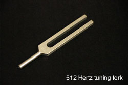
512Hz Tuning Fork

Trigeminal Nerve – Pain and Temperature

Examination Technique:

  • explain to the patient that you will be touching them with a sharp object. Reassure them that it is disposable and has not been used on anyone else.
  • for pain, use either a pin or the sharp end of a broken tongue depressor.
  • use a cold tuning fork to assess temperature. If necessary, the tuning fork can be cooled by running it under cold water.
  • ask the patient to report whether they feel sharp or dull or cold.
  • ask the patient if the two sides feel the same.

Trigeminal Nerve – Motor

The motor component of the trigeminal nerve (V3) supplies the muscles of mastication. The largest of these include the temporalis and masseter muscles.

Examination Technique:

  • palpate the temporalis and masseter muscles on either side when the patient clenches their teeth.
  • ask the patient to open their mouth and repeat this against resistance. Observe for any deviation of the jaw to one side.
  • with their mouth open, ask the patient to protrude their jaw to either side against resistance.
  • the jaw-jerk reflex is elicited by the examiner placing their index finger over the middle of the patient’s chin with the mouth slightly open and the jaw relaxed. The index finger is then tapped with a reflex hammer, delivering a downward stroke. The afferent impulse for this reflex is the sensory portion of the trigeminal nerve. The efferent limb is through the motor (V3) branch of the trigeminal nerve.

Normal Response:

  • the jaw should not deviate to either side.
  • the jaw-jerk is usually absent or weakly present.

Abnormal Response:

  • the jaw deviates towards the side of weakness.
  • the jaw-jerk is exaggerated and pathologically brisk with lesions affecting the pyramidal pathways above the 5th nerve motor nucleus, especially if the lesions are bilateral.