Ovarian Teratoma
Cystic Teratoma
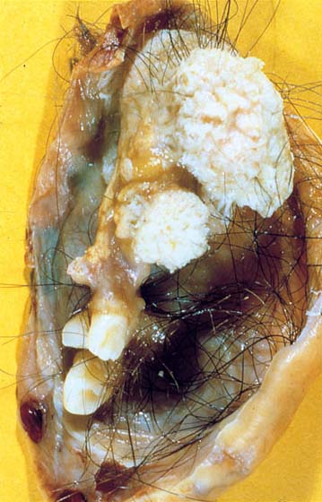
Gross appearance of an ovary with a mature cystic teratoma. (Courtesy of Dr. I. Zberiranowski)
Study Smarter
Cystic Teratoma
Gross appearance of an ovary with a mature cystic teratoma. (Courtesy of Dr. I. Zberiranowski)
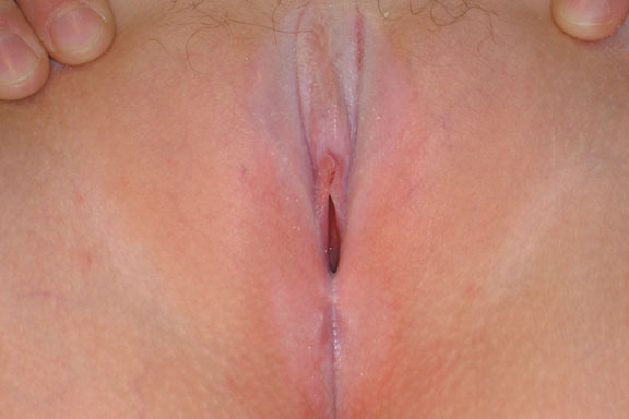
Note classic hour glass or figure 8 vulvular and perianal distribution. (Courtesy of Dr. S. Kives and Dr. R. Spitzer)
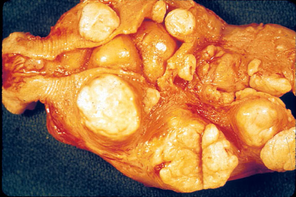
Uterus with multiple leiomyomas. (Courtesy of Dr. I. Zberiranowski)
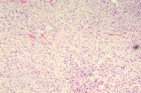
Microscopic view of proliferative smooth muscle cells. (Courtesy of Dr. I. Zberiranowski)
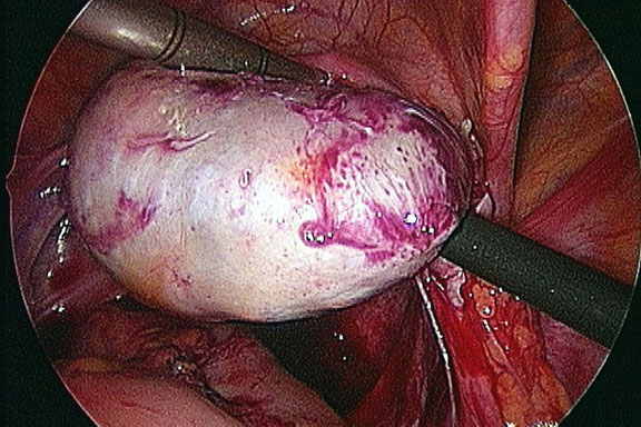
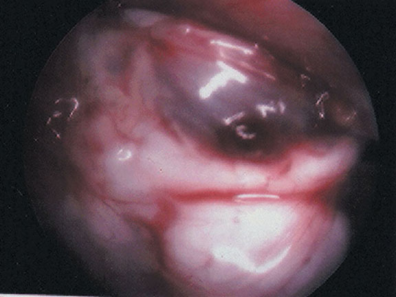
Laparoscopic Image of an Ovarian Cyst
(Courtesy of Dr. S. Kives and Dr. R. Spitzer)
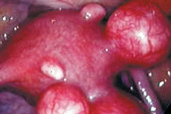
Laparoscopic Image of Multiple Fibroids
Note multiple sub-serosal fibroids.
Laparoscopic Image of Normal Female Anatomy
Note the uterus (left) and the ovary (right). (Courtesy of Dr. S. Kives and Dr. R. Spitzer)

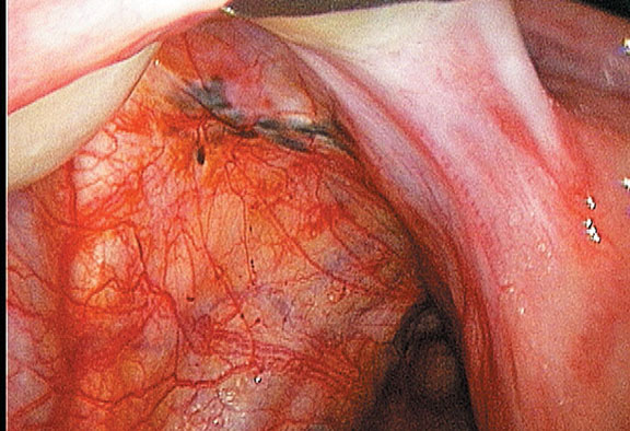
Laparoscopic Image of Endometriosis
Note brownish-black “powder burn” implants. (Courtesy of Dr. S. Kives and Dr. R. Spitzer)
Cystic mass on ovary arising from ectopic endometrial tissue. Commonly referred to as a “chocolate cyst”.
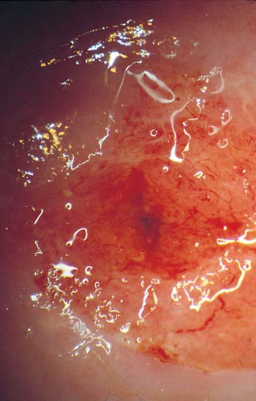
Eversion of cervical canal, with columnar epithelium farther outside the external os of the cervix. (Courtesy of Dr. G. Likrish)