Tibial Plateau Fracture
Lateral view and AP view.
Study Smarter
Lateral view and AP view.
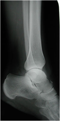
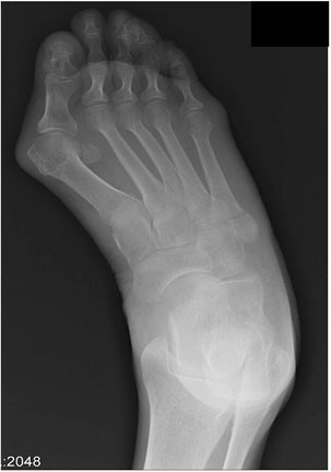
Chip fracture from talus. Note the small triangular fleck of bone lifted from the superior surface of the neck of the talus just proximal to the talo-navicular joint. This is commonly seen as a small fragment of bone with cortex all around or as a small projecting exostosis. In either case, it probably represents the appearance of an old fracture in this region.
The present film represents the appearance of the recent fracture which is not very often seen. It is probably due to pull of ligaments around the talo-navicular joint.
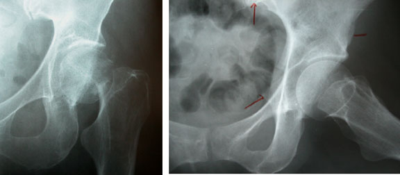
The image to the left is of a slipped capital femoral epiphysis in a child. The image to the right is after fixation. [Courtesy of Dr. Tim Dowdell, St. Michael’s Hospital, Department of Medical Imaging]
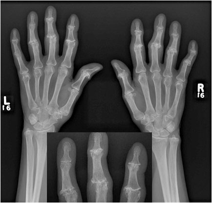
Rheumatoid arthritis erosions, joint space narrowing, and subluxations can be seen in this x-ray. [Courtesy of Dr. N. Jaffer]
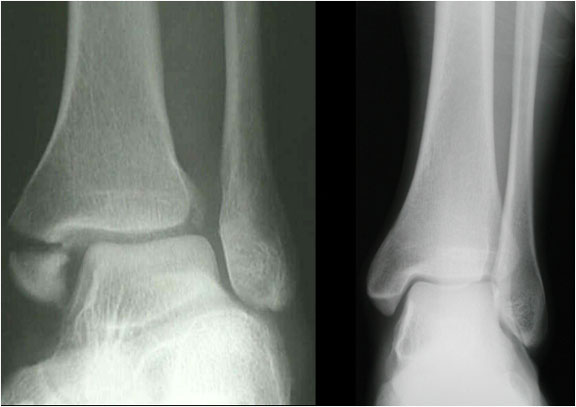
Avulsion fracture of the medial malleolus (left). Normal shown (right) for comparison.
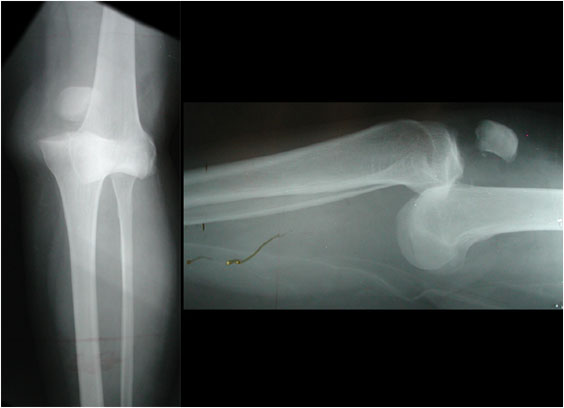
Lipohemarthrosis (left) with fracture of lateral tibial plateau (right). Lipohemarthrosis is the mixture of fat and blood in a joint cavity following trauma. Specifically, intraarticular fractures can lead to migration of fat and blood from the marrow cavity into the joint space. Due to the fact that fat is less dense than blood, a fat-blood interface forms and can be seen on radiography as a fat-fluid level.
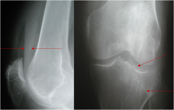
[Courtesy of Dr. Tim Dowdell, St. Michael’s Hospital, Department of Medical Imaging]
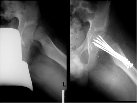
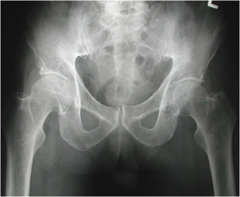
This is a single AP projection of the pelvis and hips. There is a fracture through the greater trochanter on the left side with very little displacement of fragments. Such an injury is usually produced by direct trauma. [Courtesy of Dr. Tim Dowdell, St. Michael’s Hospital, Department of Medical Imaging]
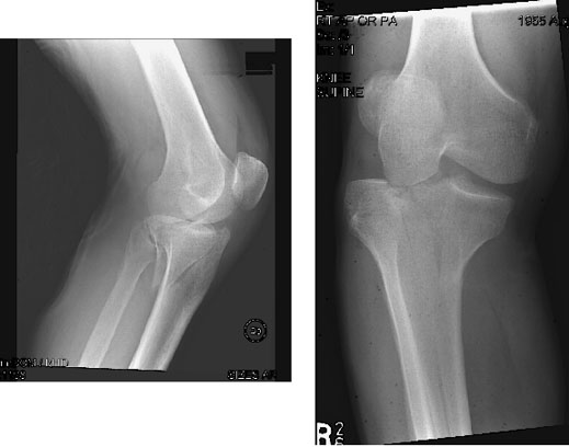
Complete dislocation of the knee. This patient suffered severe trauma in a motor vehicle accident.
The PA (left) and later (right) projections show complete dislocation of the knee with overriding and shortening.
The tibia and fibula together are anterior to the femur. The patella has retained its attachment to the tibia.
There is a small fracture from the posterior surface of the patella.