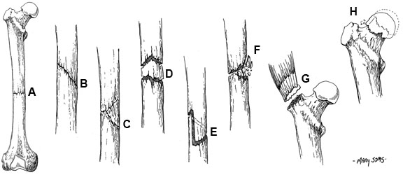
Fracture Types
A. Transverse
B. Oblique
C. Butterfly
D. Segmental
E. Spiral
F. Comminuted
G. Avulsion
H. Impacted
Study Smarter
A. Transverse
B. Oblique
C. Butterfly
D. Segmental
E. Spiral
F. Comminuted
G. Avulsion
H. Impacted
Generally you should image the limbs bilaterally (unaffected and affected), image joints immediately proximal and distal to the affected joint, take AP and lateral views (and special views depending on the joint, i.e. skyline for knee or transcapular for shoulder) and then image before and after the reduction.
Site: Identify which bone, region of bone (proximal, distal, metaphyseal, etc.), intra- or extra-articular. Look for radiolucent (dark on x-ray) lines, discontinuities in the contour of the cortex.
Type: Transverse, oblique, spiral, comminuted.
Displacement: Undisplaced or displaced (angulated, translated, rotated, shortened, impaction).
Soft Tissue involvement: Calcification, gas, foreign bodies, open vs. closed. You may see features if fracture is not obvious, such as soft tissue swelling, changes in fat stripes, joint effusions and fat fluid levels caused by the displacement of periarticular fat by joint fluid.
Joints: Are the articular surfaces in apposition? Radiolucent lines? Arthritis features?
A negative x-ray does not exclude a fracture (especially in scaphoid, radial head or metatarsal head). Diagnosis is often clinical and not confirmed until 7-10 days later when enough bone resorption has occurred.