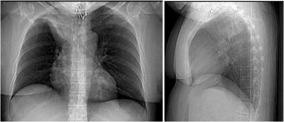
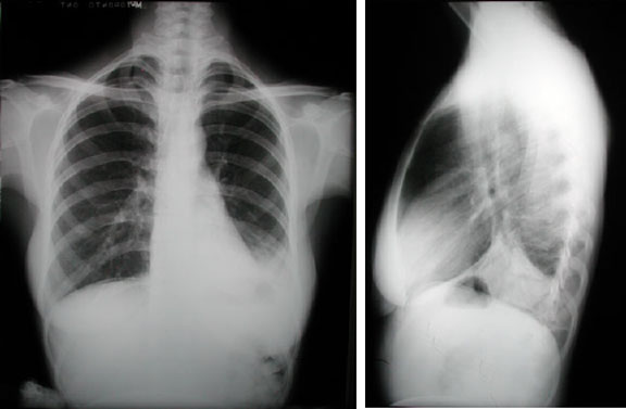
Atelectasis

RUL Collapse

This patient has suffered complete collapse of the right upper lobe. Note the opaque, atelectatic right upper lobe projecting over the superior right hemithorax. This image is a good example of the “S-sign” of Golden: The displaced minor fissure resembles an inverted “S” in contour. The collapse is most likely due to a mass in the region of the right superior lobar bronchus. The collapsed right upper lobe also demonstrates the “silhouette” sign; in this case, the adjacent superior vena cava’s border is obscured by the right upper lobe. There is mild elevation of the right hemidiaphragm, consistent with atelectasis on the right. Although not convincingly seen here, other indirect signs such as hilar and mediastinal shift can also be present with atelectasis. Multiple surgical clips are noted in the region of the thoracic inlet.

RUL Collapse

RML Collapse

This patient has suffered complete collapse of the right middle lobe. Note the opaque, atelectatic right middle lobe projecting over the mid right hemithorax. The collapsed right middle lobe also demonstrates the “silhouette” sign; in this case, part of the adjacent right heart border is obscured by the atelectatic lobe. The lateral view demonstrates the flat, “pancake”-shaped right middle lobe. The minor fissure (black arrow) and major fissure (green arrow) approximate as the intervening lung tissue collapses.

RML Collapse

RLL Collapse

This patient suffered complete collapse of the right lower lobe. The major fissure is clearly displaced inferiorly, the atelectatic lobe projects as a flat opacity, and the right hemidiaphragm is “silhouetted” (see below). Indirect signs of atelectasis include a right shift of the mediastinum. Other indirect signs such as compensatory hyperinflation, elevation of the right hemidiaphram, and hilar shift are not seen here. This study is an excellent example of the “silhouette” sign. In a normal chest x-ray, the diaphragm and mediastinal structures are visible because of the difference in radiodensity between lung and these structures (i.e there is an “interface” between the tissues). The “silhouette” sign refers to a loss of normally appearing interfaces, implying opacification due to consolidation (most common), atelectasis, mass, etc., in adjacent lung. In this case, the collapsed right lower lobe “silhouettes” the right hemidiaphragm. Note that the right heart border, adjacent to the right middle lobe, is clearly visible.

RLL Collapse

LUL Collapse

This patient has collapse of the entire left upper lobe, including the lingula. On the PA film, this projects as a hazy opacity over much of the upper left hemithorax. The lateral view shows the classic”pancake” opacity filling the retrosternal air space. There is mild elevation of the left hemidiaphragm; other indirect signs are not well seen here. This film also demonstrates “silhouetting” of the aortic knob and upper left heart border.

LUL Collapse

LLL Collapse

This patient has postoperative atelectasis of the entire left lower lobe. The left hemidiaphragm is “silhouetted” and the left hilum displaced downward. On the lateral view, there is an obvious retrocardiac density (the atelectatic lobe) and downward displacement of the fissure. This patient also has a postoperative pneumoperitoneum.

LLL Collapse